by Nancy W. Burkhart, BSDH, EDD
A new patient today, Becky, is a 44 year-old female who is taking Synthroid and ranitidine HCL 150 mg. Becky is a non-smoker, occasional alcohol user, and married with no general complaints. Her last visit to a dental office was approximately four years ago.
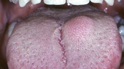
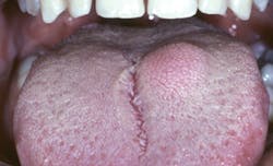
Becky informs you that she has a "bump" on the left dorsal surface of her tongue. As you begin your oral exam, you palpate this area of the tongue, and your first impression is that it is probably a fibroma. However, the firmness that usually denotes the fibroma is not present. The nodule is somewhat firm but not within the usual consistency of a fibroma. The nodule measures 1.1 centimeters.
------------------------------------------------
Other articles by Nancy Burkhart
------------------------------------------------
Becky reports that the nodule is asymptomatic, and it has been present for several months. The patient is referred to an oral surgeon. The nodule is completely removed and the diagnosis is granular cell tumor of the tongue (GCT) (see Figure 1).
Granular cell tumors, also referred to as Abrikossoff tumor, may affect any area of the body, but the head and neck are affected in up to 65% of occurrences. Of these findings, approximately 70% are found on the tongue, followed by the oral mucosa. GCT may occur in various other locations such as on the skin, the lungs, thyroid, larynx, trachea, parotid gland, etc. The tumor is considered benign, but malignancy is known to occur on rare occasions, comprising approximately 2% of the total cases. The tissue size is usually 3 mm or less, painless, a non-ulcerated nodule and presents as a solitary lesion. The GCT has a mean average of discovery in the fourth to sixth decade of life (Neville, et al. 1995), but the entity may be found at any age. There is a female predilection of 2:1. The color is usually pink, but may also have a yellow hue.
Pseudoepitheliomatous hyperplasia (irregular proliferation of the epithelium) or acanthosis may occur, and may mimic squamous cell carcinoma when the biopsy is shallow (Del'Horto, et al.2013). When this mistaken diagnosis of squamous cell carcinoma is made, it will mean unnecessary, extensive surgery for the patient. So, a careful diagnosis with confirmation of the histology and an excellent surgical procedure is crucial.
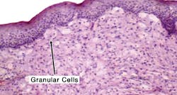
Histology: The tissue specimen consists of large polygonal cells that have granular cytoplasm containing lysosomes. The cells are separated by collagen bundles. Biopsy with histological determination is important to differentiate the tissue from that of schwannomas, neurofibroma, irritation fibromas, hemangiomas, and lymphangiomas. Immunohistochemical assay is also performed.
The malignant GCT is suggested when there is a spindle cell pattern, mitotic activity, necrosis and wide cellular sheets (Giuliani, et al. 2004) The absence of mitotic figures, hyperchromatism, increased nuclear cytoplamic ratio etc., will rule out the possibility of a malignancy. The GCT is not encapsulated, therefore the margins are important considerations in total removal (see Figure 2).
Treatment: Surgical removal is necessary to establish a diagnosis. Recurrence is not likely with complete removal. However, adequate follow-up is standard procedure.
Conclusion: Careful palpation of the tongue is a key component of the intraoral exam. Small nodules such as the GCT may be detected at an early stage. This means less surgery for the patient and an early diagnosis. Since the GCT occurs predominately on the tongue, you may be the first health-care provider to diagnose this abnormality and assist the patient in receiving adequate treatment.
As always, continue to ask good questions, and always listen to your patients. RDH
NANCY W. BURKHART, BSDH, EdD, is an adjunct associate professor in the department of periodontics, Baylor College of Dentistry and the Texas A & M Health Science Center, Dallas. Dr. Burkhart is founder and cohost of the International Oral Lichen Planus Support Group (http://bcdwp.web.tamhsc.edu/iolpdallas/) and coauthor of General and Oral Pathology for the Dental Hygienist. She was a 2006 Crest/ADHA award winner. She is a 2012 Mentor of Distinction through Philips Oral Healthcare and PennWell Corp. Her website for seminars on mucosal diseases, oral cancer, and oral pathology topics is www.nancywburkhart.com.
References:
Dell'Horto AG, Pinto JM, Diniz MS. Case for diagnosis. Abrikossoff's tumor: Unusual location. An Bras Dermatol. 2013;88(3):469-71.
Dive A, Dhobbley A, Fande PZ, Dixit S. Granular cell tumor of the tongue: Report of a case. J Oral Maxillofac Pathol. 2013. 17(1)148.
Giuliani M, Lajolo C, Pagnoni M, Boari A, Zannoni GF. Granular cell tumor of the tongue (Abrikossoff's tumor). A case report and review of the literature.
Minerva Stomatol. 2004;53:465-9.
Neville B W, Damm DD, Allen CM, Bouquot JE. Oral & Maxillofacial Pathology. 1995; W.B. Saunders Co. Philadelphia.
Dive A, Dhobley A, Fande PZ, Dixit S. Granular cell tumor of the tongue: Report of a case. J Oral Maxillofac Pathol [serial online] 2013 [cited 2013 Jul 4];17:148. Available from: http://www.jomfp.in/text.asp?2013/17/1/148/110728.
Past RDH Issues


