Case #9
by Joen Iannucci Haring
A 24-year-old female visited a dentist for evaluation of yellowish depressions on her upper teeth.
History
The patient first noticed the yellowish depressions on her upper teeth within the past year. When questioned about any change in size, the patient stated that the areas appeared to be enlarging. No history of trauma to the affected areas was reported by the patient, and the patient denied any pain or discomfort associated with the affected teeth. When questioned about dietary products, the patient denied the excessive ingestion of soft drinks or lemons.
At the time of the dental appointment, the patient appeared to be in an overall good state of health. The patient's medical history was reviewed and included no significant positive findings. The patient was not taking medications of any kind at the time of the examination.
The patient's past dental history included regular dental examinations and routine dental treatment. The patient's last dental appointment was two years earlier for a dental prophylaxis.
Examinations
Physical examination of the head and neck area revealed no enlarged or palpable lymph nodes. The patient's vital signs were obtained and all found to be within normal limits. No significant extraoral findings were noted.
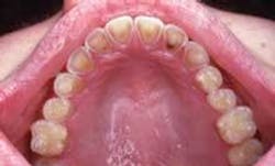
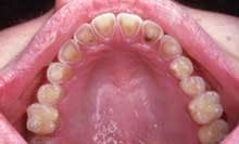
Intraoral examination revealed yellowish depressions on the lingual aspects of the maxillary teeth (see photo). The depressions appeared shallow, broad, smooth, and scooped out.
Further examination of the facial surfaces of the teeth revealed no other lesions present.
Diagnosis
Based on the clinical information presented, which one of the following is the most likely diagnosis?
o attrition
o abrasion
o erosion
o enamel hypoplasia
o fluorosis
Diagnosis
• erosion
Discussion
Erosion can be defined as the loss of tooth structure that results from the chemical action of acids. Any acid that is placed in prolonged contact with teeth produces a drop in pH that results in erosion. Bacteria are not involved, as with dental caries. The acids that cause the demineralization process may be from a dietary, external, or internal source. Dietary sources of acid include lemons and other acidic fruits, vinegar, and soft drinks. External sources include acids found in the work environment (for example, battery manufacturing).
The most common internal source of acid is from gastric secretions following regurgitation; chronic regurgitation may be associated with conditions such as bulimia, the hiatal hernia, esophagitis, or pregnancy.
Clinical features
Erosion may affect the labial, lingual, or occlusal surfaces of teeth. The lesions, which may vary in size, shape, and appearance, usually involve more than one tooth. There are different patterns of erosion that may be identified; the pattern often indicates the causative agent or the particular habit. For example, a chronic lemon-sucking habit produces characteristic changes seen on the facial surfaces of the maxillary anterior teeth. Horizontal ridges are first seen in the enamel and then followed by smooth, cupped-out, shallow, yellowish depressions in dentin adjacent to the cervical portion of the crowns. The same pattern is also seen in swimmers who chronically expose their teeth to highly chlorinated pool water.
Another pattern of erosion that may occur presents as a smooth loss of enamel with exposure of dentin on the lingual surfaces of the maxillary teeth. If the lingual surface of the tooth is involved, a concave area of dentin surrounded by a thin, white line of enamel along the periphery of is seen. Chronic vomiting is associated with these characteristic changes of the lingual surfaces of the maxillary teeth. If the posterior teeth are involved, an extensive loss of enamel results on the occlusal surfaces. In posterior teeth with amalgam restorations, the edges of the restorations may extend above the level of the remaining tooth structure. Occasionally, entire cusps may be lost to erosion.
Erosion may or may not be symptomatic. The most common symptom of erosion is sensitivity of dentin in the exposed areas. In some instances, erosion may progress and not only result in dentinal sensitivity, but in pulpal exposure as well.
Differential diagnosis
The clinical appearance of erosion is characteristic. Patient history is important to determine the causative acid or offending habit. Erosion does not resemble functional wear patterns (attrition, for example) or wear patterns associated with abnormal mechanical forces (abrasion, for example) and should not be confused with such conditions.
Treatment
Early diagnosis and intervention are necessary to prevent further tooth loss that results from erosion. Patient education can be used to modify behavior once the source of acid or offending habit is identified. In most instances, habit elimination or behavior modification is necessary for success.
To restore form and alleviate dentinal sensitivity, areas of erosion should be treated with conventional operative procedures (for example, composite resins, veneers, onlays, or crowns).
Joen Iannucci Haring, DDS, MS, is a professor of clinical dentistry, Section of Primary Care, The Ohio State University Coolege of Dentistry.

