Case #8
By Joen Iannucci Haring
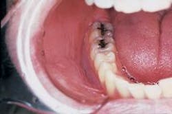
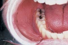
A 25-year-old male presented to a dental office for a checkup. Oral examination revealed a white, wrinkled area in the mucobuccal fold.
History
The patient was unaware of the white area inside the mouth and denied any pain or sensitivity associated with the lesion. The patient did not report any history of trauma to the affected area.
At the time of the dental appointment, the patient appeared to be in an overall good state of health. No significant problems were obtained during review of the health history, and the patient was not taking any medications at the time of the dental examination.
The patient's past dental history included regular dental examinations and routine treatment. The patient's last dental visit had taken place approximately one year earlier for a checkup and dental prophylaxis. A review of the patient's record revealed no evidence of a white lesion in the mucobuccal fold at the last dental visit.
Examinations
Physical examination of the head and neck region revealed no enlarged or palpable lymph nodes. The patient's blood pressure and vital signs were all found to be within normal limits. Extraoral examination revealed no abnormal findings.
Intraoral examination revealed a large, diffuse white area in the mucobuccal fold (see photo). The lesion appeared wrinkled or corrugated in texture. The lesion could not be removed by wiping or scraping. Further oral examination of the oral soft tissues revealed no other lesions present.
Clinical diagnosis
Based on the clinical information presented, which of the following is the most likely diagnosis?
o smokeless tobacco keratosis
o verrucous carcinoma
o proliferative verrucous leukoplakia
o frictional hyperkeratosis
o idiopathic leukoplakia
Diagnosis
• smokeless tobacco keratosis
Discussion
The term smokeless tobacco keratosis refers to an oral lesion that results from the use of a smokeless tobacco product. The development of such a lesion is related to the length of exposure to the smokeless tobacco product. There is a direct relationship between the use of smokeless tobacco and mucosal changes; exposure of the oral tissues to the tobacco product results in both chemical and cellular injury.
Smokeless tobacco products include both snuff and chewing tobacco. Snuff can be defined as a finely ground or powdered tobacco that is sold in both dry and moist forms. Snuff is not chewed; instead a small amount (referred to as a "pinch") is placed and held in the mucobuccal fold area between the cheek and gingiva, or between the lower lip and gingiva.
Tobacco sold in loose-leaf, compacted "plug" or brick form is known as chewing tobacco. As the name suggests, chewing tobacco is held in the mouth, typically inside the cheek, and chewed. While chewing tobacco most often is used by older adults, moist snuff is commonly used by teenagers.
For years, the use of snuff and chewing tobacco was traditionally limited to a small segment of the population. Previously identified as a habit of cowboys, men over the age of 65, and baseball players, smokeless tobacco products are now widely used by adolescents and young adults, especially athletes. The increase in smokeless tobacco use is believed to be the result of media advertising and peer pressure.
Clinical features
Although a smokeless tobacco keratosis is typically seen in young males, this lesion may be seen in any individual who uses a smokeless tobacco product. The lesion has a fairly characteristic clinical appearance and is found at the preferred site of tobacco placement. If more than one intraoral location is used to hold the tobacco products, multiple and less prominent lesions may be seen. The most frequent sites of involvement include the mucobuccal fold area in the incisor and molar regions.
The typical smokeless tobacco keratosis appears white with a raised or flat contour. The texture of the lesion appears smooth, granular, or corrugated. The appearance of the keratosis has been compared to that of an "ebbing" tide. A smokeless tobacco keratosis is asymptomatic and is usually discovered upon routine oral examination.
Diagnosis and treatment
The diagnosis of a smokeless tobacco keratosis can be made based upon the history of product use and the characteristic clinical appearance. Histologic examination reveals chevron-shaped projections, acanthosis, and hyperkeratosis. Dysplasia and/or squamous cell carcinoma may be seen with a history of longtime use of smokeless tobacco products.
If the use of the smokeless tobacco product is discontinued, the lesion should disappear after two to six weeks. After discontinuing the use of smokeless tobacco, all lesions that last longer than six weeks must be biopsied.
An individual with a smokeless tobacco keratosis should be advised to discontinue using the tobacco product.
In addition, the dental professional can provide such patients with information on the effects of smokeless tobacco use and problems that may result. Patients should be advised that the long-term use of smokeless tobacco products may result in oral problems that not only include the smokeless tobacco keratosis, but periodontal tissue changes, dental caries, and dental abrasion. The use of smokeless tobacco products may also cause nicotine dependence, alterations of taste and smell, and high blood pressure.
Because smokeless tobacco products contain carcinogens known at nitrosamines, longtime exposure to such products may result in malignant changes and lesions such as verrucous carcinoma or squamous cell carcinoma.
For additional information concerning smokeless tobacco and how it affects the oral cavity, dental professionals may contact the American Cancer Society at www.cancer.org or at (800) ACS-2345, or the National Oral Health Information Clearinghouse at www.nohic. nih.gov.
Joen Iannucci Haring, DDS, MS, is a professor of clinical dentistry, Section of Primary Care, The Ohio State University College of Dentistry.

