Tonsilloliths: Opacities on panoramic images
BY NANCY W. BURKHART, BSDH, EdD
Tonsilloliths are caseous masses (coagulation necrosis) or calcifications that collect in the crypts of the palatine tonsils. These may also be found in the adenoidal crypts or surrounding lymphoid tissue.
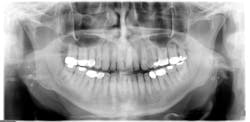
For the most part, the concretions go undetected unless they are observed in a panoramic radiograph (see Figure 1). Patients may report coughing up what is described as firm pieces of food or debris that have an odor to them. The patient may also complain of halitosis and a foul taste. In rare instances, the patient may report odynophagia (burning pain or squeezing pain when swallowing) and dyspnea (difficulty in breathing or shortness of breath). Patient may also complain of ear pain.
-------------------------------------------
Other columns by Burkhart
- Lichen planus pemphigoides: Diagnosis should be followed by referral
- Reduction of Tooth Stains: Patient education is vital to preventing tooth stain
- Oral Pemphigus and Pemphigoid: Why are these conditions so hard to diagnose?
-------------------------------------------
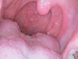
Unless the patient brings this to the attention of a health-care provider, the enlargement of the tonsil may go unnoticed until the patient is examined during an oral cancer exam or the calcifications are noticed on a panoramic radiograph. The importance of examining the tonsillar region should be emphasized not only because of entities such as tonsillitis, but also because of tonsillar cancer or other oral cancers affecting the posterior region of the mouth (see Figure 2).
The Concretions: The concretions may be white or yellow and present in various shapes as small pellets. They collect in enlarged tonsillar crypts or surrounding lymphoid tissue. They are sometimes initiated when inflammation occurs such as an infection of the throat or ear. Chronic inflammation may cause enlargement of the crypts and subsequent fibrosis.
The debris forms over time within the crypts and eventually may calcify. Balaji et al. (2013) describe the concretions as consisting of organic debris, bacteria, calcium salts, calcium carbonate apatite, oxalates, and other magnesium salts and ammonium radicals. The environment of the tonsillar crypts promotes anaerobic bacterial progression. The saliva adds the needed components to make calcification possible. Others report that the debris may also contain fungus and actinomycosis as well (Lo, 2011). Increased amounts of sulfur compounds produce the putrid odor that is often detected by the patient and the health-care provider.
Giant tonsilloliths are not common, but when very large, they may be mistaken for pathology when viewed on a radiograph. Careful clinical assessment is needed.
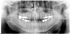
Radiographic assessment: Small, multiple, radiopaque, ill-defined, calcareous objects are seen on the radiograph (see Figure 1). The radiograph presented in this column was taken in 2015 of a 76-year-old male.
These opacities are not limited to the palatine tonsils but may also be seen in accessory lymphoid tissue. Sometimes what is referred to as "ghost images" appear on a pantomograph, making a pseudotonsillolith visible on the opposite side. This occurs because the object is located between the X-ray source and the center of rotation of the cassette. The patient may have oral calcifications bilaterally or unilaterally, so further evaluation is always needed.
Differential diagnosis: Diagnosis is made by radiographic assessment of the location exhibiting the oral calcifications. Calcifications may be found in other areas of the body such as within the molar-ramus region, including sialoliths, phleboliths, calcified lymph nodes, carotid artery arteriosclerosis, stylohyoid ligament ossification, and dystrophic calcification in acne scars (Bamgbose et al., 2014) Calcifications can also occur throughout the body - not just oral tissues.
Epidemiology: A study (Bamgbose et al., 2014) reported statistics on a group of patients presenting with tonsilloliths and other calcifications. Out of 124 selected cases of tonsillitis, 53 were males and 71 were females with a male-to-female ratio of 0.75:1.00. The age range was from 9.2 to 87 years old with a mean age of 52.6 years of age. The average size of the tonsillolith was 4 mm with a range of 3-11 mm. Other study data appear to indicate that these entities may occur in any age group with a purported average of 46.2 years of age.
Treatment: Most tonsilloliths are annoying to the patient, but they are usually small and pose no problems. However, when infections such as ear pain or chronic sore throat, abscesses, dysphagia, or halitosis are a problem for the patient, surgical removal is usually the option.
Concretions can be removed by using a water irrigator to dislodge some of the debris. The patient would lean over a sink so that the dislodged material would flow into the basin. These patients have such deep crypts that it is virtually impossible to remove all the material and only surface areas are reached. In rare cases, the patient may have such tonsil enlargement due to the tonsilloliths that the airway blockage becomes a concern.
Conclusion: The tonsillar region and posterior region of the mouth are important areas to examine during the oral cancer exam. A patient may have the concerns discussed within this column, but they often do not attribute any issues or complaints to the tonsillar region. Tonsillar cancer begins within the tonsillar crypts and behind the tonsillar tissue. Assessing this area through visual, tactile, and radiographic interpretation is so important. The dental lights and reflective mirrors make many tonsilloliths visible to the practitioner and can be demonstrated to the patient during a dental visit.
As always, continue to ask good questions and always listen to your patients. RDH
References
Balaji BB, Avinash Tejasvi ML, Anulekha Avinash CK, Chittaranjan B. Tonsillolith: A panoramic radiograph presentation. JCDR. 2013; Oct, 7:10: 2378-2379.
DOI:10.7860/JCDR/2013/5613.3530.
Bamgbose BO, Ruprecht A, Hellstein J, Timmons S, Qian F. The prevalence of tonsilloliths and other soft tissue calcifications in patients attending oral and maxillofacial radiology clinic of the University of Iowa. ISRN Dentistry. 2014, Article ID 839635. http://dx.doi.org/10.1155/2014/839635.
Dal Rio AC, Franchi-Teixeira AR, Nicola EMD. Relationship between the presence of tonsilloliths and halitosis in patients with chronic caseous
tonsillitis. BDJ 2007;204:E4. DOI: 10.1038/bdj.2007.1106.
Guevara C, Mandel L. Panoramic radiographic demonstration of bilateral tonsilloliths. NYSDJ. 2011 Apr; 77(3):28-30.
Lo RH, Chang KP, Chu ST. Upper airway obstruction caused by bilateral giant tonsilloliths. J Chin Med Assoc. 2011; 74(7): 329-31.
Oda M, Kito S, Tanaka T, et al. Prevalence and imaging characteristics of detectable tonsilloliths on 482 pairs of consecutive CT and panoramic radiographs. BMC Oral Health 2013; 13:54. doi:10.1186/1472-6831-13-54.
Ram S, Siar CH, Ismail SM, Prepageran N. Pseudo bilateral tonsilloliths: a case report and review of the literature. Oral Surg Oral Med Oral Pathol Oral Radiol Endod. 2004 July;98(1):110-4.
NANCY W. BURKHART, BSDH, EdD, is an adjunct associate professor in the department of periodontics, Baylor College of Dentistry and the Texas A & M Health Science Center, Dallas. Dr. Burkhart is founder and cohost of the International Oral Lichen Planus Support Group (http://bcdwp.web.tamhsc.edu/iolpdallas/) and coauthor of General and Oral Pathology for the Dental Hygienist. She was a 2006 Crest/ADHA award winner. She is a 2012 Mentor of Distinction through Philips Oral Healthcare and PennWell Corp. Her website for seminars on mucosal diseases, oral cancer, and oral pathology topics is www.nancywburkhart.com.


