Targets, techniques, and technology help ensure compliance with screening protocols
By Nicole Giesey, RDH, BSDH, MSPTE
In my 10 years of practicing dental hygiene, I have developed a certain routine. While every patient is different and treatment can vary, my core routine has been in place until recently. I'm now including the opportunity for every patient to have an advanced oral cancer screening taken with the newest technology on the market. This is in addition to the traditional visual/tactile oral cancer screening that I've incorporated in my routine since hygiene school. I have the extreme pleasure of working in a practice that has incorporated the newest technology in daily practice, and this technology includes the Identafi oral cancer screening instrument. I've used ViziLite and VELscope in the past, and I was eager to learn this new technology. In other offices that had oral cancer screening technology, I found, oddly, that it was rarely used. It was almost as if the doctor had a new toy that was initially used; then it slowly faded as the newer toy came along, or the doctor decided to use it only on patients he felt fit a certain criteria.
RISKS FOR ORAL CANCER
Tobacco/Alcohol Use
HPV
Age
Sun Exposure
Diet
The responsibility to ensure the screenings are being performed lies with the hygienist. This is yet another task to complete in the limited time we have. I have found that if the hygienist is not inspired by the technology, patients do not receive the benefit of the technology. The question I pose to hygienists is: Are you fitting an oral cancer screening into your appointments? Perhaps you're using the technology only on certain people. Does oral cancer have a certain criteria? According to the Oral Cancer Foundation, it is estimated that 40,000 people in the United States will be diagnosed with oral cancer or pharyngeal cancer this year (Oral Cancer Foundation, 2013).
Who do you think should receive an oral cancer screening? The answer is easy. Every adult and every young adult, especially those who fall into the high-risk category, should receive a screening. What are the risk factors? According to the Mayo Clinic, they are tobacco use of any kind, heavy alcohol use, excessive sun exposure to the lips, and a sexually transmitted virus called human papillomavirus (Mayo Clinic, 2012). Even though some patients are outside of the risk factor criteria for oral cancer, they should still have the opportunity to benefit from a screening. Early detection is key to beating oral cancer, and as with any cancer, the earlier the detection, the higher the survivor rate.
The Oral Cancer Foundation stated, "The death rate for oral cancer is higher than that of cancers which we hear about routinely such as cervical cancer, Hodgkin's lymphoma, laryngeal cancer, cancer of the testes, and endocrine system cancers such as thyroid, or skin cancer (malignant melanoma)" (OCF, 2013). If this is the statistic, and we are encouraging women to get pap smears yearly and men to have testicular cancer screenings, then we as hygienists need to make it a point to incorporate oral cancer screenings into our routine. If your dentist is already performing an oral cancer screening and you feel that one exam is enough, just follow the old saying that two sets of eyes are better than one. How often have you found faulty restorations or decay that your dentist missed, simply because you're in the patient's mouth a lot longer than the dentist is? The patient can only benefit from your set of trained eyes and professional tactile exam.
How often are oral cancer screenings recommended? The American Dental Association recommends that an oral cancer screening should be completed at the periodic oral exam visit (American Dental Association, 2012). Does this mean that every prophy with a periodic exam needs to have the advanced technology oral cancer screening, or is the visual/tactile oral cancer screening sufficient? This is up to the practice. Our office recommends the visual/tactile screening every periodic exam, and an advanced screening in conjunction with the visual/tactile screening once a year. This way, at every periodic exam, the patient receives a screening of some sort. Need to brush up on your visual/tactile exam methods? There are excellent resources available at your fingertips.
SIGNS OF ORAL CANCER
A sore, irritation, lump or thick patch in the mouth, lip, or throat
A white or red patch in the mouth
A feeling that something is caught in the throat
Difficulty chewing or swallowing
Difficulty moving the jaw or tongue
Numbness in the tongue or other areas of the mouth
Swelling of the jaw that causes dentures to fit poorly or become uncomfortable
Pain in one ear without hearing loss
Whenever I'm writing to my fellow hygienists, I like to include avenues of information that are helpful in our daily hygiene life. One very useful publication comes from the National Oral and Craniofacial Research Institute and is titled "Detecting Oral Cancer: A guide for health care professionals." This oral cancer screening guide can be found on their website at http://www.nidcr.nih.gov/OralHealth/Topics/OralCancer/DetectingOralCancer.htm. It is the best brochure on the topic that I've found, and it is free!
This brochure is not only a great tool for the dental hygiene student, but it is a wonderful addition to even the most experienced hygienist's library. This brochure shows both photographically and in writing the step-by-step methodology of the oral cancer visual/tactile examination. Patient education information and brochures can also be found on this website. The patient education information brochures are a great chairside conversation piece while waiting for an examination. Other publications about oral cancer from the American Dental Association can be ordered on the site. There is an abundance of printable patient education available online if you don't want to order brochures. A list of other websites with valuable information are:
- http://oralcancerfoundation.org/
- http://www.mayoclinic.com/health/mouth-cancer/DS01089
- http://www.ada.org
The visual/tactile examination should never be replaced by the technology examinations. The technology can be used in addition to the visual/tactile examination, which is extremely important in the early detection of oral cancer. The correlation to HPV is important to us as hygienists so we can ensure that we're examining the posterior areas of the oral cavity where the HPV associated lesions occur. A thorough examination of the entire oral cavity is not painful to the patient and does not take a lot of time. There are three technologies on the market that I will describe and discuss. These are ViziLite, VELscope, and Identafi.
If you've never used these technologies, their resources are only a click away. A quick visit to their websites will give you an idea of their technology and how it works. Some sites also have video tutorials. To check out the VELscope technology, visit http://www.velscope.com/. Click on "Dental Professionals" for their video. To learn about ViziLite, visit http://www.zila.com/. This website is very nice, but there is no video. The Identafi website (http://www.identafi.net/) provides a great training video and research information. If visiting the websites is not enough and your office is interested in incorporating one of these technologies into the practice, call the company for a lunch and learn. Although the technologies are all used for the same purpose, they are all different in design and usage.
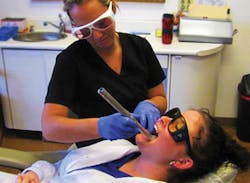
The differences between the technologies are vast. ViziLite incorporates a 1% acetic acid rinse prior to examination with a light. If a lesion appears during examination and there is not a history of trauma, a swab of acetic acid is applied around the lesion. Following the acetic acid is a swab of TBlue dye for a more extensive examination (Zila, 2012). The VELscope and Identafi are different from the ViziLite in that there are no rinses or swabs, only lights. The VELscope technology uses a blue light that enables the clinician to see any suspicious areas that show up dark under the light. This technology is used in a dark room. VELscope also has a camera accessory to use for documentation purposes if any suspicious area is present (VELscope, 2012). The VELscope looks just like its name suggests – a scope or flashlight. The Identafi technology is similar to VELscope with its blue light, and it has two additional lights to assist in detecting suspicious tissue vasculature. The additional lights are an amber light to tell the difference in vasculature of normal and suspicious tissue, and a white light (Identafi, 2012). The Identafi is used with disposable mirror heads. This technology is also used in a darkened room, and both the patient and clinician wear protective eyewear.
Even if your office does not currently use these technologies, you never know when you'll use them in the future. A quick read or video on them will help you understand how they work and their differences. Check with your state’s laws and regulations on hygienists’ use of technology and performing the adjunctive prediagnostic screening. In some states, dental hygienists are not legally able to complete this procedure. I dream of the day all states are united in a universal dental hygiene law to benefit patients nationwide.
There is a CDT procedure code to use when completing this exam – D0431. It is described as an adjunctive prediagnostic screening. Many insurance companies do not cover this code, but incorporating it into your routine and educating the patient on the importance of early detection would easily justify the low cost of the exam. Please do not let insurance companies dictate your care. At your next meeting, a team discussion regarding the office's oral cancer screening protocol could invite a new standard of care. How many of the 40,000 new cases will you detect early with your screenings?
When you or the dentist completes an oral cancer screening, proper documentation is a must. There are mouth maps available on standard dental chart templates, or you can use a mouth map document provided from one of the technology websites. If you are paperless, most dental software has an oral exam screen with descriptor words already formatted for you. When documenting a lesion either on paper or computer, be sure to include size, shape, color, surface texture, consistency, location, and previous trauma (Wilkins, 2009). The dental probe really comes in handy when measuring lesions. If your doctor wants to reevaluate the patient, document in the patient’s chart that the dentist recommends having the lesion rechecked in two weeks. If it’s still present after two weeks, your doctor will most likely refer the patient to an oral surgeon. Document the recommendation with a copy of the referral in the patient's chart.
Now that you’ve properly examined, educated, and documented the oral cancer screening, you’re part of the oral cancer fighting team. We need to be consistent in our oral cancer screening endeavors. All patients need our professional and trained eyes. If we continually raise the bar on our standard of care, our futures can only be brighter. A universal bar needs to be raised in the fight against oral and pharyngeal cancer. How will you help raise it? RDH
Consider reading: Oral cancer awareness: Do good in the world. Do good in your practice.
http://www.dentistryiq.com/articles/2013/03/oral-cancer-awareness-do-good-in-the-world-do-good-in-your-pract.html
Consider reading: Discussing the horrors of oral cancer with two survivors
http://www.dentistryiq.com/articles/2012/09/discussing-the-horrors-of-oral-cancer.html
Consider reading: Dental Health Promotion: Giving oral cancer a louder voice
http://www.rdhmag.com/articles/print/volume-32/issue-10/features/giving-oral-cancer-a-louder-voice.html
References
American Dental Association. (2012). Cancer, Oral. http://www.ada.org/2607.aspx
Dental EZ group. (2012). Identafi. http://www. identafi.net/
LED Medical Diagnostics Inc. (2013). VELscope. http://www.velscope.com/
Mayo Foundation for Medical Education and Research. (2012). Mouth Cancer. http://www.mayoclinic.com/health/mouth-cancer/DS01089
Oral Cancer Foundation. (2013). Oral Cancer Facts. http://oralcancerfoundation.org/
Wilkins EM. (2009). Clinical practice of the dental hygienist (10th ed., pp. 183-90). Baltimore: Lippincott Williams & Wilkins, a Wolters Kluwer business.
Zila, a Tolmar Company. (2013). ViziLite. http://www.zila.com/40/
Nicole Giesey, RDH, BSDH, MSPTE, practices clinically part-time. She is an adjunct dental hygiene clinical instructor at Youngstown State University and a writer. She enjoys researching and writing and creating fun puzzles about dental hygiene topics. Nicole can be reached at [email protected].
Past RDH Issues


