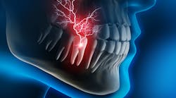
The issue of dentinal hypersensitivity has plagued clinicians and their patients since the beginning of the dental profession. While some cases are easy to diagnose and treat, others are more mysterious and complex. It’s not unusual for affected patients to return to the office, visit after visit, reporting ongoing symptoms that cannot be resolved despite their best efforts. In some of these scenarios, there is no visible radiographic or clinical pathology, which can leave the provider feeling bewildered as to what they should recommend for relief. Some contributing factors have been widely established and recognized, while others are relatively new developments in research. When approaching the topic of sensitivity, it is important to consider both the conventional and contemporary philosophies on diagnosis and treatment to provide the best individualized care for patients in need.
Related reading:
Winning the battle against sensitivity
The untold secrets of managing dentin hypersensitivity: Part 1 of 2
FluoroCal by Bisco: A great option for relieving dentinal hypersensitivity
Prevalence
Although dentinal hypersensitivity has been studied for decades, researchers have had difficulty identifying a clear proportion of the population who suffer from it. In a meta-analysis published in 2018, it was established that somewhere between 1.3% and 92.1% of subjects reported experiencing hypersensitivity.1 This broad range was said to be influenced by participant age, study size, and participant recruitment method. An average obtained from all studies showed that about 33.5% of participants reported symptoms consistent with dentinal hypersensitivity. While the percentage remains slightly unclear, what studies do tell us is that hypersensitivity in individuals is not an unusual occurrence. Most clinicians would agree with this notion, as it can be presumed that many encounter a fair number of patients suffering from hypersensitivity during their careers.
The physics of hypersensitivity
Many theories have been developed to explain the phenomenon of dentinal hypersensitivity, from a network of nerves present in the dentin to the concept of odontoblasts sensing pain in the pulpodentinal border.2 Many of these theories have been rejected upon further investigation, with microscopic evaluation disproving the idea that nerve tissue extends to the outer cells of the tooth. The hydrodynamic theory, on the other hand, has been more widely recognized by researchers and is currently taught to students by educators in clinical programs. This concept suggests that fluid flowing in the dentinal tubules stimulates a nociceptor response from the pulp and dentin. According to this theory, teeth that have wider and/or more numerous tubules have an increased likelihood of experiencing hypersensitivity. For this reason, many treatment options are aimed at either occluding the tubules or desensitizing sensory nerves to alleviate the patient’s symptoms.
Causes
Even though the science behind sensitive teeth seems simple, there are many contributing—sometimes complicated—factors that can lead to a person’s struggle with hypersensitivity. Many of these elements have been known in the dental profession for decades, while some have been more recently investigated and proven. Having knowledge of all possible reasons a patient is experiencing hypersensitivity aids in identifying the most appropriate solution.
Exposed dentin
Whether it’s caused by gingival recession, tooth abrasion and/or abfraction, cracked enamel, acid erosion, decay, or a fractured tooth, dentin is bound to be sensitive when it is exposed to extreme temperatures and heavy occlusal forces without the natural protection from healthy enamel and gingival tissue. While exposed dentin doesn’t guarantee hypersensitivity symptoms, it significantly increases the likelihood. Patients can experience hypersensitivity in specific, localized areas due to exposed dentin, or in mouth-wide, generalized cases, depending on the number of lesions and the severity of their involvement.
Teeth whitening
There are two primary ways to whiten teeth: physical removal of stains and chemical lightening of stains and enamel. In the case of physical stain removal, these whitening products have an abrasive property that detaches stain particles from the tooth surface. In some cases, these abrasive kinds of toothpaste and gels not only eliminate stains, but also gradually damage the gingival tissue and tooth structure exposed, leading to sensitivity. Chemical whitening, on the other hand, bleaches and degrades chromogens by way of either carbamide or hydrogen peroxide. The oxidation process that occurs with bleaching products can effectively lighten stained teeth, while also opening occluded tubules in the dentin, which leads to sensitivity.3 Certain whitening products have both physical stain removal and bleaching properties, increasing the risk of causing sensitivity. Product strength and frequency of use can also affect sensitivity symptoms.
Periodontal disease and therapy
The inflammatory process that results in bone reduction, tooth loss, and a wide array of potential systemic ailments4 can also cause hypersensitivity due to an absence of protective gingiva and bone structure.5 Aside from the disease itself, surgical and nonsurgical supportive therapy can also inadvertently increase symptoms of sensitivity due to subsequent exposure of root surfaces during surgical excision of tissue and removal of insulating layers of calculus deposits, as well as minor removal of cementum during scaling and root-planing instrumentation. In many cases, patients experience more postoperative discomfort with external stimuli than they did prior to treatment.
Iatrogenesis
The primary goal of restorative dentistry is to protect and preserve an individual’s dentition, thereby improving their health, self-confidence, and overall quality of life. Many dental procedures utilize a combination of technique-sensitive clinical skills, advanced specialized knowledge, and a variety of restorative materials that are designed to simulate natural tooth structure and function. Even with the clinician’s best efforts and intentions, sometimes complications arise after dental treatment that can produce hypersensitivity in the patient. Changes in thermal insulation, occlusion, pulpal trauma, open margins between the restoration and the tooth structure, improper fit, defective material, or errors in the execution of the procedure can ultimately result in either temporary or long-term sensitivity for the patient.
Bruxism
The act of clenching or grinding the teeth together on a frequent basis can result in a variety of negative effects on the patient, from temporomandibular joint (TMJ) pain, muscular pain, headaches, structural damage to the teeth, and dentinal hypersensitivity. It is important to realize that cases of bruxism differ from person to person; not every patient shares the same symptoms, frequency, or reason for their habit. From a clinical aspect, signs of bruxism typically present as incisal or occlusal wear patterns, cervical lesions on the tooth, gingival recession, chipped or broken teeth, mobility, or the presence of tori.6
Emerging research suggests that current events such as the COVID-19 pandemic have increased and amplified the population’s bruxing habits while awake, as emotional stress and mental illness can serve as triggers for clenching and grinding.7 Another study performed in 2021 found that a worldwide increase in search engine inquiries about bruxism occurred after the onset of the pandemic.8 In addition to stress-induced bruxism, mounting evidence indicates a relationship between nighttime bruxing and sleep-disordered breathing, such as obstructive sleep apnea.9 Individuals who experience frequent sleep arousals due to interrupted breathing have a high risk of sleep bruxism and should be treated accordingly.
Solutions
There are a variety of possible treatment options available for a patient who is experiencing chronic hypersensitivity. It is important to fully investigate the true source of the individual’s symptoms to ensure the problem is being properly managed or eliminated. In the case of topical sensitivity relief, a study was done in 2019 that determined that:10
Glutaraldehyde with HEMA, glass ionomer cements, and laser present significant immediate (until 7 days) dentinal hypersensitivity reduction. Medium-term (until 1 month) reduction was observed in stannous fluoride, glutaraldehyde with HEMA, hydroxyapatite, glass ionomer cements, and laser groups. Finally, long-term significant reduction was seen in potassium nitrate, arginine, glutaraldehyde with HEMA, hydroxyapatite, adhesive systems, glass ionomer cements, and laser.
Essentially, the study communicated that each of these topical options has a purpose and a place in the therapeutic relief of dentinal hypersensitivity, depending on how quickly relief is needed and for how long. The decision lies with the clinician and patient as they agree on the approach that will best suit the patient’s individual needs.
Aside from desensitization and tubule occlusion, evaluating the patient’s dentition for iatrogenic sources of hypersensitivity is also a worthwhile effort. If found, these areas will likely need to be restoratively retreated or, in some cases, referred to the appropriate collaborating specialist for management. Patients tend to be highly appreciative and trusting of providers who acknowledge a restorative issue and assist them in correcting it to improve their function and level of comfort.
When considering hypersensitive patients who show signs of bruxism, discussing the nature of their bruxing habit is of utmost importance in order to provide the most appropriate treatment option. As mentioned, obstructive sleep apnea can be highly linked to bruxism, and although the two conditions are not dependent upon each other, screening a bruxing patient for sleep-disordered breathing before solely focusing on their clenching or grinding is wise. Recent research indicates that approximately 20% of American adults suffer from obstructive sleep apnea, with 90% of them being undiagnosed.11 Asking a patient who bruxes simple questions about their quality of sleep, snoring, and daytime sleepiness can help guide the provider in the right direction. Clinically evaluating the patient for signs of airway obstruction, such as scalloped tongue, narrow airway, large tonsils, mouth-breathing, or retrognathia can aid in the clinician’s discernment of the situation. If these concerns are seen, referral to the patient’s primary care physician for a sleep study is warranted. Many dental offices that are centric on sleep-disordered breathing utilize pulse oximetry or polysomnography devices to monitor their patients’ sleep health and collaborate with a physician to treat the patient’s bruxism and sleep concerns. If the screening process yields no concerns for the patient’s airway health, addressing their bruxism can be accomplished through the fabrication of an occlusal guard, stress reduction techniques, counseling on sleep hygiene, and behavior modification.
Dentinal hypersensitivity is a multifaceted problem that affects many people. Discomfort can stem from a wide range of sources and can, in turn, require various options for treatment and relief. Having knowledge of these contributors and their solutions will help the oral health-care provider ensure they are treating their patients as functionally and progressively as possible. Although it seems that many people suffer from dentinal hypersensitivity at some point in their lives, we must remember that each patient in our operatory has a unique history, perception of pain, and desired outcome. Individualized care is paramount in providing relief for patients in need.
Editor's note: This article appeared in the August 2022 print edition of RDH magazine. Dental hygienists in North America are eligible for a complimentary print subscription. Sign up here.
References
- Favaro Zeola L, Soares PV, Cunha-Cruz J. Prevalence of dentin hypersensitivity: Systematic review and meta-analysis. J Dent. 2019;81:1-6. doi:10.1016/j.jdent.2018.12.015
- Langenbach F, Naujoks C, Smeets R, et al. Scaffold-free microtissues: differences from monolayer cultures and their potential in bone tissue engineering. Clin Oral Investig. 2013;17(1):9-17. doi:10.1007/s00784-012-0763-8
- Carey CM. Tooth whitening: what we now know. J Evid Based Dent Pract. 2014;(14 Suppl):70-76. doi:10.1016/j.jebdp.2014.02.006
- Hegde R, Awan KH. Effects of periodontal disease on systemic health. Dis Mon. 2019;65(6):185-192. doi:10.1016/j.disamonth.2018.09.011
- Lin YH, Gillam DG. The prevalence of root sensitivity following periodontal therapy: a systematic review. Int J Dent. 2012;2012:407023. doi:10.1155/2012/407023
- Bertazzo-Silveira E, Stuginski-Barbosa J, Porporatti AL, et al. Association between signs and symptoms of bruxism and presence of tori: a systematic review. Clin Oral Investig. 2017;21(9):2789-2799. doi:10.1007/s00784-017-2081-7
- Hassan KA, Khier SE. Awake bruxism intensified during COVID-19 pandemic by cumulative stress: an overview. J Clin Res. 2020;3(1):1-3. doi:10.33309/2639-8281.030103.
- Kardes E, Kardes S. Google searches for bruxism, teeth grinding, and teeth clenching during the COVID-19 pandemic. J Orofac Orthop. 2021;1-6. doi:10.1007/s00056-021-00315-0
- Hosoya H, Kitaura H, Hashimoto T, et al. Relationship between sleep bruxism and sleep respiratory events in patients with obstructive sleep apnea syndrome. Sleep Breath. 2014;18(4):837-844. doi:10.1007/s11325-014-0953-5
- Marto CM, Baptista Paula A, Nunes T, et al. Evaluation of the efficacy of dentin hypersensitivity treatments-a systematic review and follow-up analysis. J Oral Rehabil. 2019;46(10):952-990. doi:10.1111/joor.12842
- Finkel KJ, Searleman AC, Tymkew H, et al. Prevalence of undiagnosed obstructive sleep apnea among adult surgical patients in an academic medical center. Sleep Med. 2009;10(7):753-758. doi:10.1016/j.sleep.2008.08.007


