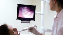
Multiple applications for the images have evolved for the hygiene operatory
BY Jamie Collins, RDH, CDA, RDH
In my practice, the intraoral camera holds a place of importance right up there near the X-ray unit and my loupes. The value it can provide in many ways is essential to any practice. The use of intraoral cameras in daily practice can serve multiple purposes from patient education to attaching images with insurance claims.
Image quality and ease of use of intraoral cameras have improved tremendously over the years. Most are now very slim and ergonomically designed handpieces with a single button for capturing images quickly, while others may require the click on a computer screen's icon to capture the desired images. Most are corded and can plug into the USB port in the computer. As with many dental products, multiple manufacturers of intraoral cameras are able to bridge with different radiography software. Similar to computer programs and products, each intraoral camera system has its own features, and some are compatible to specific software. Most dealer reps will offer a demonstration of the various camera types that are compatible to an office's software systems.
What do they really see?
As clinicians, we use magnification to enable us to see better, but what about our patients? We try to explain what we see or try to show them in a hand mirror. But can they really see what we see? Usually the patient doesn't see the same things we see; they may just trust but not fully understand or visualize what we are trying to explain. I have had many patients to whom I have tried to educate and explain the need for periodontal therapy and the significant amount of calculus present, only to have the patient request a "regular cleaning."
Once I show the patient a large-screen photo of their bleeding gingiva and inflammation in addition to the calculus present, they are suddenly more interested in their oral condition and usually are more responsive to treatment recommendations. Many patients are then able to grasp an understanding of the condition and are often willing to complete the recommended treatment.
As a proponent for patient education, I always like to explain the reasoning behind any treatment recommendations that are made in the practice. By using intraoral imaging, I am able to show as well as tell the patient what I see. How many times have patients put off what they can't see because it is not hurting at the time? With the use of intraoral imaging, the patient sees the broken filling or recurrent decay on the computer screen, and most do not want that in their mouths. Due to the increased use of intraoral cameras over the years, I have seen case acceptance for treatment increase greatly. I even have patients ask me to take an image so they are able to see the problem as well. I used to rarely use the intraoral camera while explaining an oral condition, but then discovered that imaging and education complemented each other in the dental practice.
What insurers see too
Many individuals want to do only treatment that dental insurance will help pay for. One great way to aid in the preauthorization process is to provide an intraoral image to the insurance carrier.
An example is when a patient has a tooth that needs a crown due to fracture lines and a large filling. The radiograph does not show a fracture, and may or may not show the decay depending on the surface of the tooth. The patient may state nothing hurts and want to know if insurance will pay a portion of the proposed treatment before they decide to proceed. Take an intraoral image and show the patient the fractures. A good intraoral image will show the fracture lines in clear detail.
Submitting the intraoral image along with the preauthorization or insurance claim may help the approval process of needed treatments such as crowns and even periodontal therapy.
'Watching' disease progress
We see hundreds of patients, and we can't remember every mouth every time. Clinically, we complete the periodontal charting to monitor pocket depth and recession. We refer to and compare the numbers at each visit. Dentists and hygienists see multiple patients daily, and often find areas to "watch" for a period of time, including incipient decay, soft-tissue lesions, or other concerns.
One fantastic way to monitor these areas is with an intraoral image to refer back to at each subsequent dental visit. Having the ability to compare photographs side by side to evaluate and monitor changes establishes a standard of care that is essential in any dental practice. The technology in the operatory also shows patients that the office is current with technology to provide the best care possible.
I have at least one patient weekly who questions how a restoration or other condition compares to the previous recall visit. I am able to provide the visual comparison to review with the individual, and the dentist is able to make a recommendation accordingly. The truth is that I can't remember from six months previously what the restoration on No. 14 looked like on any particular patient. The saved images help tremendously for comparison.
Setting the stage for new patients
My office allows extra time for new-patient visits for initial records, radiographs, and periodontal charting. In addition to those records, I take a series of intraoral images of existing restorations and any areas I suspect will need future treatment or monitoring, such as caries, failing restorations, abfractions, and the general gingival condition.
By doing so, I am able to review the patient's concerns in addition to any recommended treatment. Going over the intraoral images with the patient prior to the dentist coming in for an exam plants the seed within the patient's mind that there may be work to complete at future visits. It provides me with the opportunity to answer any questions I can prior to the dentist coming into the room. In turn, I am able to relay concerns to the dentist to help mainstream the exam and keep everyone on schedule.
The saying "A picture is worth a thousand words" is so true. In dentistry, it couldn't be more accurate in regard to the use of intraoral cameras. Using images to explain needed treatment helps patients visualize oral conditions as well as the possibilities to improve their smiles. Intraoral imaging is not only used to identify fractured teeth or periodontal conditions, it can also be used for areas of cosmetic improvement.
Intraoral images are necessary prior to orthodontic treatment as part of the records process in conjunction with impressions. Images can also be used to show the shade of the teeth pre- and post- whitening treatment. It can often be used to send images to the lab when cosmetic restorations are done to match shades or shape of the teeth.
When I first learned to use a mirror intraorally, it took a little time to get the hang of it; the use of the intraoral camera is very similar. There is a learning curve to adapting the camera to capture the image while trying to keep the lens fog free. I got the hang of it quickly, and the intraoral camera soon became one of the most valuable tools I have, especially for patient education and case presentation. It is essential for daily use in clinical practice and has the potential to increase production with increased case acceptance. RDH
Jamie Collins, RDH, CDA, resides in Idaho with her husband, Cory, and their four children. She currently works as a full-time hygienist as well as an educator at the College of Western Idaho. In addition, she acts as a content expert and contributor in multiple upcoming textbooks. She can be contacted at [email protected].


