Mitochondrial dysfunction, periodontitis, and systemic disease: Part 2
Editor's note: This is part two of a two-part series. Read part one here.
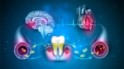
Welcome back! The role of mitochondrial dysfunction in the association between periodontitis and systemic diseases has garnered increasing attention over the past decade. Periodontal tissue, like other bodily tissues, relies on mitochondrial function for the energy production necessary for maintenance and repair. Impairment of their function can compromise the integrity and health of tissues. Let’s look at how common disease mechanisms influence mitochondria, periodontitis, and a few systemic conditions—namely, obesity, cardiovascular disease, diabetes, and cognitive impairment.
Obesity
Obesity, characterized by excessive body fat accumulation, is closely associated with periodontitis incidence due to impaired immune response, chronic inflammation, and oxidative stress (OS), which in turn affect mitochondrial function.1 Obese individuals often exhibit higher levels of pro-inflammatory factors and lower levels of anti-inflammatory factors, both systemically and locally. Periodontitis does the same.
Pro-inflammatory cytokines such as TNF-α and IL-1 can induce mitochondrial dysfunction by increasing reactive oxygen species (ROS) production and altering mitochondrial dynamics. Chronic inflammation can lead to sustained OS, causing persistent mitochondrial damage and impairing cellular energy metabolism. Studies have shown elevated periodontal OS in obese patients, which may not be significantly improved by nonsurgical periodontal therapy alone but can be effectively reduced when combined with diet therapy.2
There may be limited research on how periodontitis affects obesity, but it is suggested that Porphyromonas gingivalis (Pg) and its virulence factors, such as lipopolysaccharides (LPS), trigger adipocyte apoptosis by disrupting mitochondrial function.3
Cardiovascular disease
Normal mitochondrial function is fundamental for a healthy cardiovascular system. Mitochondria need to divide (a process called fission) to maintain quality control and cell balance, and in cardiovascular diseases (CVD) this process is often overly active.4 Proper mitochondrial function is also crucial for preventing heart aging.5
Keep in mind that periodontal patients are in a general inflammatory state, and increased ROS from neutrophils can lead to a higher likelihood of developing CVD.6 Pg can worsen atherosclerosis by damaging mitochondria.7 Pg can cause mitochondrial division and dysfunction in blood vessel cells by increasing the activity of a protein called dynamin-related protein 1 (Drp1), whose activity is tightly regulated to clear damaged mitochondria through mitophagy, keeping precise control over cellular processes and the dynamics of the heart.
There’s some good news: blocking this process with a Drp1 inhibitor called a mitochondrial division inhibitor (mdivi-1) can prevent the harmful effects of Pg, and it has been studied as a potential therapeutic not only in CVD but in neurodegenerative disorders.8 Besides division, mitochondria also influence periodontitis and CVD through OS. A component of Pg called dihydroceramide triggers inflammation and increases ROS and mitochondrial-dependent cell death in blood vessel cells. In patients with periodontitis, higher OS levels in certain blood cells may lead to atherosclerosis and other heart-related problems.9
Diabetes
There are shared pathological processes between periodontal disease (PD) and type 2 diabetes (T2D) such as OS and mitochondrial dysfunction.10 We know that bacteria such as Pg stimulate the immune response leading to ROS production, and the excess causes oxidative tissue damage. In T2D, hyperglycemia also leads to increased production of ROS through autoxidation of glucose and the formation of advanced glycation end products (AGEs), the harmful compounds that form when proteins or fats combine with sugars in the bloodstream through a process called glycation.
AGEs are also linked to the development of chronic periodontitis (CP). Experiments have demonstrated that AGEs induce the production of ROS in periodontal ligament cells, which in turn triggers autophagy.11,12 AGEs induce endogenic ROS and autophagy, activating a pathway that initiates mitochondrial-mediated apoptosis. Excess ROS contributes to insulin resistance and beta-cell dysfunction, features of T2D. Research has shown patients with CP and T2D had more bone loss and periodontal cell apoptosis than in the CP group alone, along with more severe mitochondrial dysfunction.13 Unbalanced OS emerged as a link between T2D and CP.
Cognitive impairment
Neurons depend almost entirely on mitochondria to generate the energy needed for crucial cellular processes, such as synaptic plasticity and neurotransmitter synthesis. The brain is especially susceptible to OS and damage due to its high oxygen consumption, limited antioxidant defenses, and abundant polyunsaturated fats, which are highly prone to oxidation. Pg contributes to neuroinflammation and neurodegenerative diseases due to its virulence factor-LPS triggering OS.14 Experiments showed that treating human neuroblastoma cells with Pg-LPS negatively affects mitochondrial function through toll-like receptor 4 (TLR4) activation. TRL4 is an essential activator of the innate immune response.
The accumulation of methylmalonic acid (MMA) in tissues is connected with disruptions in mitochondrial energy balance and ATP production, making it an indicator of mitochondrial dysfunction. A study found that MMA levels in the blood partly explained the link between PD indicators, such as pocket depth and attachment loss, and cognitive function, indicating that mitochondrial dysfunction connects periodontitis and cognitive decline in the elderly.15
Although periodontitis is triggered by bacteria colonizing the tooth surface and gingival sulcus, the host’s response plays a crucial role in the destruction of connective tissue and bone. Among these responses, mitochondrial function undergoes rapid and significant changes, sometimes even before the onset of inflammation. These fluctuations in mitochondrial function can be seen as critical signaling events and are often among the first indicators when cells encounter external stimuli. Diverse manifestations of mitochondrial dysfunction are observed in the various tissues and cells involved in the development and progression of periodontitis.
Mitochondrial dysfunction is a serious pathological mechanism connecting periodontitis with various systemic diseases. Bacteremias resulting from pathogens and their virulence factors entering the bloodstream can cause mitochondrial dysfunction in distant target cells, promoting the development of systemic diseases. Conversely, systemic diseases that induce OS can worsen local periodontal damage. Understanding these mechanisms and incorporating mitochondrial-targeted therapies alongside conventional mechanical debridement hold promise as innovative approaches for controlling periodontitis.
Editor's note: This article appeared in the August/September 2024 print edition of RDH magazine. Dental hygienists in North America are eligible for a complimentary print subscription. Sign up here.
References
- Zhao P, Xu A, Leung WK. Obesity, bone loss, and periodontitis: the interlink. Biomolecules. 2022;12(7):865. doi:10.3390/biom12070865
- Martínez-Herrera M, Abad-Jiménez Z, Silvestre FJ, et al. Effect of non-surgical periodontal treatment on oxidative stress markers in leukocytes and their interaction with the endothelium in obese subjects with periodontitis: a pilot study. J Clin Med. 2020;9(7):2117. doi:10.3390/jcm9072117
- Singh SP, Huck O, Abraham NG, Amar S. Kavain reduces Porphyromonas gingivalis-induced adipocyte inflammation: role of PGC-1 signaling. J Immunol. 2018;201(5):1491-1499. doi:10.4049/jimmunol.1800321
- Quiles JM, Gustafsson ÅB. The role of mitochondrial fission in cardiovascular health and disease. Nat Rev Cardiol. 2022;19(11):723-736. doi:10.1038/s41569-022-00703-y
- Picca A, Mankowski RT, Burman JL, et al. Mitochondrial quality control mechanisms as molecular targets in cardiac ageing. Nat Rev Cardiol. 2018;15(9):543-554. doi:10.1038/s41569-018-0059-z
- Sanz M, Del Castillo AM, Jepsen S, et al. Periodontitis and cardiovascular diseases. Consensus report. Glob Heart. 2020;15(1):1. doi:10.5334/gh.400
- Xu T, Dong Q, Luo Y, et al. Porphyromonas gingivalis infection promotes mitochondrial dysfunction through Drp1-dependent mitochondrial fission in endothelial cells. Int J Oral Sci. 2021;13(1):28. doi:10.1038/s41368-021-00134-4. Erratum in: Int J Oral Sci. 2022;14(1):3. doi:10.1038/s41368-021-00153-1
- Ruiz A, Alberdi E, Matute C. Mitochondrial division inhibitor 1 (mdivi-1) protects neurons against excitotoxicity through the modulation of mitochondrial function and intracellular Ca2+ signaling. Front Mol Neurosci. 2018;11:3. doi:10.3389/fnmol.2018.00003
- Zahlten J, Riep B, Nichols FC, et al. Porphyromonas gingivalis dihydroceramides induce apoptosis in endothelial cells. J Dent Res. 2007;86(7):635-640. doi:10.1177/154405910708600710
- Ferreira IL, Costa S, Moraes BJ, et al. Mitochondrial and redox changes in periodontitis and type 2 diabetes human blood mononuclear cells. Antioxidants (Basel). 2023;12(2):226. doi:10.3390/antiox12020226
- Mei YM, Li L, Wang XQ, et al. AGEs induces apoptosis and autophagy via reactive oxygen species in human periodontal ligament cells. J Cell Biochem. 2020;121(8-9):3764-3779. doi:10.1002/jcb.29499
- Fang H, Yang K, Tang P, et al. Glycosylation end products mediate damage and apoptosis of periodontal ligament stem cells induced by the JNK-mitochondrial pathway. Aging (Albany NY). 2020;12(13):12850-12868. doi:10.18632/aging.103304
- Li X, Sun X, Zhang X, et al. Enhanced oxidative damage and Nrf2 downregulation contribute to the aggravation of periodontitis by diabetes mellitus. Oxidative Med Cell Longev. 2018;2018:9421019. doi:10.1155/2018/9421019
- Verma A, Azhar G, Zhang X, et al. P. gingivalis-LPS induces mitochondrial dysfunction mediated by neuroinflammation through oxidative stress. Int J Mol Sci. 2023;24(2):950. doi:10.3390/ijms24020950
- Li A, Du M, Chen Y, et al. Periodontitis and cognitive impairment in older adults: the mediating role of mitochondrial dysfunction. J Periodontol. 2022;93(9):1302-1313. doi:10.1002/JPER.21-0620
About the Author

Anne O. Rice, BS, RDH, CDP, FAAOSH
Anne O. Rice, BS, RDH, CDP, FAAOSH, founded Oral Systemic Seminars after over 35 years of clinical practice and is passionate about educating the community on modifiable risk factors for dementia and their relationship to dentistry. She is a certified dementia practitioner, a longevity specialist, a fellow with AAOSH, and has consulted for Weill Cornell Alzheimer’s Prevention Clinic, FAU, and Atria Institute. Reach out to Anne at anneorice.com.

