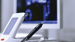
There is a greater concern than ever about aerosols, microbes, and what dental professionals can do to help protect our patients and ourselves in a post-COVID world. Here's a look at the numerous benefits of preprocedural dental laser decontamination to reduce bacteria in the sulcus, which prevents the start of inflammation and subsequent tissue destruction. We will also explore how a laser can be used postprocedurally to kill bacteria deeper than the reach of current instrumentation, stimulate fibroblasts and osteoblasts (i.e., bone-forming cells) to regenerate tissue, and trigger the periodontal cells to promote faster healing.
Diode lasers
One of the most popular lasers in dentistry is the diode laser. Several advantages of these lasers include extreme compactness, affordability, ease of operation, simple setup, and versatility. Diode lasers are particularly useful in hygiene practice because their wavelengths are highly absorbed in the melanin and hemoglobin found in the soft tissue.1 When patients don’t clean subgingivally and prolong their dental cleanings, biofilm communities increase. Healthy pink tissue becomes red in color (because it has more pigment, or melanin) and is associated with increased bleeding.
This is an opportunity for the laser to perform optimally because it can be taken into the sulcus, and at certain settings, the laser will only be attracted to and interact with diseased tissue (i.e., complex red-orange bacteria) and less strongly with healthy tissue.2 Due to the wide acceptance of the diode laser, the research referenced in this article will specifically relate to diode wavelengths, their benefits, and how easily dental hygienists can incorporate diode lasers into practice.
Why diode lasers for dental hygiene?
To understand how a laser is used, we need to look at the basics of what the laser does and why we should choose it over other tools we have in our armamentarium.
One of every two American adults aged 30 and over has periodontal disease.3 Most of our patients have some form of periodontal disease—from gingival inflammation and bleeding gums, to detachment of collagen fibers and alveolar bone loss.4
The first component of gingivitis and periodontal disease is biofilm.5 Biofilm starts out as single, free-floating bacteria in the sulcus. When it is not removed properly, these bacterial cells aggregate and bond to form a vicious gang of mature biofilm. The alarming problem with biofilm is that bacteria can move from the tooth surface and penetrate the lining of the sulcus epithelium to a depth of 500 microns. Biofilm can also get into the bloodstream and circulate throughout the body.6
The body responds to this biofilm invasion by sending fluid to the affected area to flush out the infection. Circulation becomes stagnant, swelling occurs, and metabolic products become backlogged. This process is how the gum tissue becomes puffy, inflamed, bulbous, and darker in color. The ferocious gang of bacteria in the biofilm causes a chronic wound, with some of the free-floating bacteria dispersing and starting a new gang in a nearby pocket. The destructive process continues throughout the entire mouth, moving from tooth to tooth.
Lasers can be used to eliminate the biofilm both before and after dental treatment. When done properly, preprocedural laser use can reduce the single, free-floating bacteria, in turn preventing the destruction process. When the biofilm has invaded the gum pocket, the diode laser can be used postprocedurally to collect the diseased biofilm and stimulate the tissue to heal and regenerate.
Laser bacterial reduction
Lasers work by directly affecting the biofilm and supporting the body’s healing response.2 These days, we and our patients are more cognizant of aerosols and infection control. The laser can be used as a preprocedural decontamination tool to reduce bacteria in the sulcus before we could potentially elicit bleeding on probing (BOP) or during instrumentation.7 The laser procedure is performed with an uninitiated tip and is known as laser bacterial reduction (LBR).
One of the main reasons to use a diode laser is for its bactericidal capabilities. Moritz et al.7 demonstrated the bactericidal properties of diode lasers in their study in which half of the patients were treated with a diode laser six times over a six-month period, and the other half of the study group was treated with H₂O₂ (hydrogen peroxide) six times over a six-month period. The diode laser group showed a 100% reduction in long-term bacteria, whereas just 58.4% of the controls showed an improvement. The diode laser group reduced BOP by 96.9%, compared to 66.7% in the control group.
The long-term bactericidal effect of the diode laser was remarkable on specific bacteria found in periodontal disease: Aggregatibacter actinomycetemcomitans, Prevotella intermedia, and Porphyromonas gingivalis. A study by Fontana et al.8 showed considerable bacterial elimination of Prevotella intermedia, Streptococcus bacillus, Fusobacterium sp., and Pseudomonas sp. when the diode laser was used on 40 rats induced with periodontal disease. The laser is an effective tool to reduce bacteria, specifically periodontal bacteria in the sulcus.
Performing LBR prior to a dental prophylaxis is similar to a preprocedural rinse, but instead of reducing the bacteria in the oral cavity alone, the laser reduces bacteria in the sulcus where dental hygienists take their instruments. The laser helps lower the microbial count in aerosols that are released during ultrasonic instrumentation, reduce or eliminate the risk of bacteremia, and prevent cross contamination with preventive instruments.9
Bacteremia (bacteria in the bloodstream) is a well-known side effect of ultrasonic scaling.10–17 When the tip of the ultrasonic scaler is inserted between the tooth and gingiva, the junctional epithelium and periodontal ligaments rupture, allowing bacteria from the sulcus to enter the bloodstream and cause transient bacteremia.
Assaf et al.9 found that preprocedural subgingival diode laser application reduces the incidence of bacteremia associated with ultrasonic scaling. This study also suggested that for the elimination of bacteria, lasers are superior to antibiotics in mechanism of action and local use. Since antibiotic prophylaxis is not 100% effective against all strains of oral bacteria, laser therapy might be a novel protective method for use with high-risk patients.9 Dental professionals still use antibiotics before prophylaxis if recommended by a high-risk patient’s physician, yet the use of the diode laser in conjunction with antibiotic premedication decreases the risk of bacteremia.
The same concept holds true if we limit the bacteria in the sulcus before inserting the ultrasonic tip. The microbial count in the aerosols during ultrasonic instrumentation will be reduced.9 Dangerous aerosols can remain in the operatory air for 30 minutes to two hours after a procedure.18 Reducing the potential risk aerosols produce is important not only for patients, but for the entire dental team. Our patients are hyperaware that particles can float in the air and can get into the body. Because of this, they may be more open to hearing about options for the additional procedures we can offer to decrease bacteria and other microbes present in aerosols.
Now is the perfect time to implement LBR with every adult patient in the dental hygiene chair. If the dental hygienist can perform LBR to reduce single, free-floating bacteria in the sulcus, they could stop the destruction process at step one. This would prevent the mature biofilm from forming, which would inhibit the destructive sequences of periodontal disease, and in turn limit the loss of attachment of collagen fibers and alveolar bone around the teeth. We would be protecting our patients from the risk of bacteria in the bloodstream. In addition, LBR therapy would decrease the microbial content in aerosols and prevent the spread of disease.
Laser decontamination or laser-assisted periodontal therapy
The diode laser can also be incorporated into hygiene practice postprocedurally. As a supplement to conventional methods that address tooth structure, the laser can be used to treat biofilm in the tissue wall in more diseased pockets. Just as conventional root debridement removes biofilm and accretions from the hard tooth surface, laser decontamination removes biofilm within the necrotic tissue of the pocket wall.
The laser’s energy interacts strongly with inflamed tissue components (i.e., diseased tissue and complex red-orange bacteria) and less strongly with healthy tissue.2 This nonsurgical therapy uses very low laser settings and decontaminates, rather than cuts, the tissue.19 This procedure is called laser decontamination (LD) or laser-assisted periodontal therapy (LAPT). The laser tip can be either uninitiated or initiated prior to performing this procedure, but a high-volume evacuation system should definitely be present near the working area. If the laser is used properly, on extremely low settings, it will only be attracted to and interact with darker-pigmented bacteria; it will not affect the healthy pink tissue.
Research shows that using a laser as an adjunct to scaling and root planing (SRP) produces more evident results and bacterial reduction when compared to SRP alone.20–23 Multiple studies show lasers are able to reduce periodontal pathogens in the sulcus and allow the pocket to recolonize with healthy bacterial flora.23–25
Fenol et al.23 looked at specific periodontal pathogens in a study in which each patient had one site tested with SRP alone and another with SRP plus laser. Each site was tested three times: before the procedure, after two weeks, and after two months. The test site where the laser was used as adjunctive therapy showed a significantly higher reduction in periodontal pathogens after two months, compared to SRP alone.
Kusek et al.25 followed 70 nonsmoking patients with probing depths ≥5 mm with bleeding. SRP plus the diode laser were used in 2,103 pockets. After five years, 80% of the pockets treated with a diode laser had reduced to a healthy pocket depth of ≤3 mm. Incorporating a laser into every SRP appointment is an effective way to improve clinical indices, reduce gingival bleeding and probing depths, and stimulate periodontal tissue for improved attachment.
Biostimulation
Lasers have fascinating biostimulation properties, as they have been shown to increase collagen production, enzyme activity, micro- and lymph circulation, and fibroblast and osteoblast proliferation.26,27 Diode lasers can be used in dental hygiene to help heal gum pockets, and they have been shown to assist in growing back bone.
Ren et al.26 completed a systematic review of the literature regarding in vitro studies that focused on the effects of the diode laser on fibroblasts in human periodontal tissue. The study’s findings showed a positive effect on the proliferation of both gingival fibroblasts and periodontal ligament fibroblasts, as well as their responses to inflammation.
Huertas et al.27 showed a biostimulatory effect on bone tissue as well as enhanced osteoblastic proliferation using similar laser settings as those in dental hygiene practice. Incorporating diode lasers into periodontal treatment can stimulate fibroblasts and collagen fibers, increasing periodontal tissue attachment, and stimulate osteoblasts to re-form lost bone in vertical and horizontal dimensions. This improves the overall outcome of periodontal therapy procedures.
Laser safety and training
Prior to performing laser procedures on live patients, it is recommended that clinicians complete hands-on training for competency. Some states have requirements regarding laser use and training for dental hygienists and dentists, as explained in the state’s practice act. Along with competency training, safety training is critical for all clinicians who plan to utilize a laser. Each office owning a laser needs a laser safety officer (LSO) who is responsible for administering a safety program and making sure the dental team is educated in the safe use of the laser.28 The LSO’s duties include, but are not limited to, enforcing safety practices, providing “laser in use” signs to place outside rooms where the units are used, making sure the laser is turned to “off” or “standby” when not in operation, and providing laser wavelength-specific safety glasses to all clinicians and patients in the nominal ocular hazard distance (NOHD). The LSO also encourages use of a high-volume evacuation system and high-filtration mask (level 3 or N95), limits reflective surfaces during all laser procedures, tests each laser daily, and keeps records of incidents of laser failure or any adverse effects related to laser therapy.2,28
There are multiple ways lasers can be incorporated into dental hygiene practice from the standpoint of prevention: treating bacteria and infection; providing biostimulation of fibroblasts and osteoblasts; and healing the soft tissue and periodontal pockets. Now is the time to bring this message to the forefront of the dental community, as we provide hygiene care to patients who have a heightened sense of awareness regarding the microbial content of aerosols, cleanliness, and aerosol management in the wake of COVID-19. This will allow patients to see the benefits of laser technology and give our dental colleagues the opportunity to understand laser technology, settings, techniques, and verbiage to incorporate into each dental hygiene visit.
Editor's note: Originally posted in 2020 and updated regularly
References
- Verma SK, Maheshwari S, Singh RK, Chaudhari PK. Laser in dentistry: an innovative tool in modern dental practice. Natl J Maxillofac Surg. 2012;3(2):124–132. doi:10.4103/0975-5950.111342
- Convissar RA. Principles and Practice of Laser Dentistry. New York, NY: Mosby; 2011.
- Eke PI, Dye BA, Wei L, et al. Prevalence of periodontitis in adults in the United States: 2009 and 2010. J Dent Res. 2012;91(10):914-920. doi:10.1177/0022034512457373
- Armitage GC. Clinical evaluation of periodontal diseases. Periodontol 2000. 1995;7:39-53. doi:10.1111/j.1600-0757.1995.tb00035.x
- Fux CA, Costerton JW, Stewart PS, Stoodley P. Survival strategies of infection biofilms. Trends Microbiol. 2005;13(1):34-40. doi:10.1016/j.tim.2004.11.010
- Manor A, Lebendiger M, Shiffer A, Tovel H. Bacterial invasion of periodontal issues in advanced periodontitis in humans. J Periodontol. 1984;55(10):567-573. doi:10.1902/jop.1984.55.10.567
- Moritz A, Schoop U, Goharkhay K, et al. Treatment of periodontal pockets with a diode laser. Lasers Surg Med. 1998;22(5):302-311. doi:10.1002/(sici)1096-9101(1998)22:5<302::aid-lsm7>3.0.co;2-t
- Fontana CR, Kurachi C, Mendonça CR, Bagnato VS. Microbial reduction in periodontal pockets under exposition of a medium power diode laser: an experimental study in rats. Lasers Surg Med. 2004;35(4):263-268. doi:10.1002/lsm.20039
- Assaf M, Yilmaz S, Kuru B, Ipci SD, Noyun U, Kadir T. Effect of the diode laser on bacteremia associated with dental ultrasonic scaling: a clinical and microbiological study. Photomed Laser Surg. 2007;25(4):250-256. doi:10.1089/pho.2006.2067
- Lofthus JE, Waki MY, Jolkovsky DL, et al. Bacteremia following subgingival irrigation and scaling and root planing. J Periodontol. 1991;62(10):602-607. doi:10.1902/jop.1991.62.10.602
- Waki MY, Jolkovsky DL, Otomo-Corgel J, et al. Effects of subgingival irrigation on bacteremia following scaling and root planing. J Periodontol. 1990;61(7):405-411. doi:10.1902/jop.1990.61.7.405
- Fine DH, Korik I, Furgang D, Myers R, Olshan A, Barnett ML, Vincent J. Assessing pre-procedural subgingival irrigation and rinsing with an antiseptic mouthrinse to reduce bacteremia. J Am Dent Assoc. 1996;127(5):641-642,645-646. doi:10.14219/jada.archive.1996.0276
- Daly CG, Mitchell DH, Highfield JE, Grossberg DE, Stewart D. Bacteremia due to periodontal probing: a clinical and microbiological investigation. J Periodontol. 2001;72(2):210-214. doi:10.1902/jop.2001.72.2.210
- Dajani AS, Taubert KA, Wilson W, et al. Prevention of bacterial endocarditis: recommendations by the American Heart Association. J Am Dent Assoc. 1997;128(8):1142-1151. doi:10.14219/jada.archive.1997.0375
- Baltch AL, Schaffer C, Hammer MC, et al. Bacteremia following dental cleaning in patients with and without penicillin prophylaxis. Am Heart J. 1982;104(6):1335-1339. doi:10.1016/0002-8703(82)90164-8
- Reinhardt RA, Bolton RW, Hlava G. Effect of nonsterile versus sterile water irrigation with ultrasonic scaling on postoperative bacteremias. J Periodontol. 1982;53(2):96-100. doi:10.1902/jop.1982.53.2.96
- Kinane DF, Riggio MP, Walker KF, MacKenzie D, Shearer B. Bacteraemia following periodontal procedures. J Clin Periodontol. 2005;32(7):708-713. doi:10.1111/j.1600-051X.2005.00741.x
- Baumann K, Boyce M, Catapano-Martinez D. Transmission precautions for dental aerosols. Decis Dent. 2018;4(12):30-32,35.
- Coluzzi DJ, Convissar RA. Atlas of Laser Applications in Dentistry. Chicago, IL: Quintessence; 2007.
- Crispino A, Figliuzzi MM, Iovane C, et al. Effectiveness of a diode laser in addition to non-surgical periodontal therapy: study of intervention. Ann Stomatol (Roma). 2015;6(1):15-20.
- Elavarasu S, Suthanthiran T, Thangavelu A, Mohandas L, Selvaraj S, Saravanan J. LASER curettage as adjunct to SRP, compared to SRP alone, in patients with periodontitis and controlled type 2 diabetes mellitus: a comparative clinical study. J Pharm Bioallied Sci. 2015;7(suppl 2):S636-S642. doi:10.4103/0975-7406.163579
- Gupta SK, Sawhney A, Jain G, et al. An evaluation of diode laser as an adjunct to scaling and root planning in the nonsurgical treatment of chronic periodontitis: a clinico-microbiological study. Dent Med Res. 2016;4(2):44-49.
- Fenol A, Boban NC, Jayachandran P, Shereef M, Balakrishnan B, Lakshmi P. A qualitative analysis of periodontal pathogens in chronic periodontitis patients after nonsurgical periodontal therapy with and without diode laser disinfection using benzoyl-DL arginine-2-naphthylamide test: a randomized clinical trial. Contemp Clin Dent. 2018;9(3):382-387. doi:10.4103/ccd.ccd_116_18
- Moritz A, Gutknecht N, Doertbudak O, et al. Bacterial reduction in periodontal pockets through irradiation with a diode laser: a pilot study. J Clin Laser Med Surg. 1997;15(1):33-37. doi:10.1089/clm.1997.15.33
- Kusek ER, Kusek AJ, Kusek EA. Five-year retrospective study of laser-assisted periodontal therapy. Gen Dent. 2012;60(6):540-543.
- Ren C, McGrath C, Jin L, Zhang C, Yang Y. Effect of diode low-level lasers on fibroblasts derived from human periodontal tissue: a systematic review of in vitro studies. Lasers Med Sci. 2016;31(7):1493-1510. doi:10.1007/s10103-016-2026-4
- Huertas RM, De Luna-Bertos E, Ramos-Torrecillas J, Leyva FM, Ruiz C, García-Martínez O. Effect and clinical implications of the low-energy diode laser on bone cell proliferation. Biol Res Nurs. 2014;16(2):191-196. doi:10.1177/1099800413482695
- Laser Institute of America. ANSI Z 136.3—Safe Use of Lasers in Health Care. Florida: Laser Institute of America; 2018.
About the Author
Joy Raskie, RDH
Joy Raskie, RDH, is the CEO of Advanced Dental Hygiene, specializing in hands-on dental laser education. As a world-renowned lecturer, she conducts live and online laser training courses as well as in-office consulting. She has been practicing as an RDH in Colorado for 17 years. Her passion for lasers led her to obtain a fellowship and two associate fellowships through World Clinical Laser Institute and advanced proficiency in dental lasers from the Academy of Laser Dentistry. Raskie’s goal is to boost excitement and confidence in incorporating dental lasers into daily practice. For questions or laser training, visit advanceddentalhygiene.com or email [email protected].

