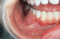
A 14-year-old male visited a general dentist for evaluation of a persistent bony swelling of the lower jaw.
Joen Iannucci Haring, DDS, MS
History
The patient first noticed the bony swelling of the lower jaw several months earlier. No pain or paresthesia was associated with the involved area. The patient appeared to be in a general good state of health with no significant medical history. The patient`s dental history included regular dental examinations and routine dental treatment. At the time of the dental appointment, the patient was not taking medications of any kind.
Examinations
The patient`s vital signs were all found to be within normal limits. Examination of the head and neck region revealed no enlarged or palpable lymph nodes. Intraoral examination revealed a localized bony hard swelling buccal to teeth #28, 29, and 30 (see photo). The mass measured approximately 2.5 cm in diameter. Further examination of the oral tissues revealed no other lesions present.
Radiographic findings
Following the intraoral examination, a periapical radiograph of the involved area was exposed. Examination of the radiograph revealed a large, well-defined unilocular radiolucency extending from the mesial of tooth #28 to the distal of tooth #30 (see radiograph).
Clinical diagnosis
Based on the clinical and radiographic information available, which of the following is the most likely diagnosis?
- central giant cell granuloma
- odontogenic myxoma
- odontogenic keratocyst
- aneurysmal bone cyst
- ameloblastoma
Diagnosis
_ central giant cell granuloma
Discussion
The central giant cell granuloma (CGCG) is an uncommon lesion found within the bones of the jaws. As its name suggests, central refers to the location of the lesion within bone, and giant cell refers to the unique giant cells that are evident upon histologic examination. The cause of this benign lesion is uncertain.
Clinical features
The CGCG is seen in all age groups, although the vast majority of cases occur in persons under the age of 30. Females are affected far more frequently than males (2:1). The CGCG occurs as a solitary lesion with 70 percent of cases occurring in the mandible. The area anterior to the first molar is most often involved and mandibular lesions frequently cross the midline.
The CGCG is an asymptomatic, slow-growing and expansile bony hard lesion. Occasionally, pain and paresthesia are features. Facial asymmetry may be noted as the lesion enlarges.
Radiographic features
On a dental radiograph, the CGCG appears as a well-defined radiolucency that is usually multilocular, although it may also appear unilocular. The size of this lesion is variable and ranges from 1 cm to 10 cm. The CGCG cannot be diagnosed from its radiographic appearance alone; a biopsy is necessary.
Diagnosis
In order to establish a definitive diagnosis, a biopsy and histologic examination of the lesion is required. Histologi-cally, the CGCG exhibits multinucleated giant cells in a background of highly cellular vascular stroma. Other lesions that may be considered in the differential diagnosis for the CGCG include the ameloblastoma (RDH April 1990), odontogenic myxoma, odontogenic keratocyst (RDH June 1989) and aneurysmal bone cyst.
Treatment
Treatment for the CGCG is surgical removal. Following the complete surgical removal, the prognosis is good. If removal of the CGCG is incomplete, the lesion may recur; recurrence rates range from 11 percent to 50 percent. Careful radiographic follow-up is essential.
Joen Iannucci Haring, DDS, MS, is an associate professor of clinical dentistry, Section of Primary Care, The Ohio State University College of Dentistry.


