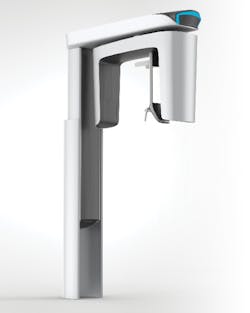
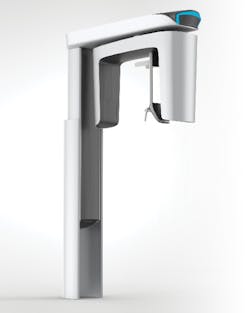
CBCT
Once unimaginable, the benefits of cone-beam computed tomography continue to increase
Ann-Marie DePalma, RDH, MEd, FADIA, FAADH
From practice management software, to digital impression scanning, to digital radiographic imaging, technology is what patients are expecting, and practices can use it for growth. Even with digital radiographic imaging modalities, such as wired sensors or photostimulable phosphor plates, the dental professional is still only viewing a three-dimensional item in two dimensions, as with film-based (analog) imaging. Since Wilhelm Conrad Röntgen’s discovery of x-rays in 1895, dental imaging has changed minimally—with a faster speed of film to decrease patient exposure and with the advent of digital dentistry.1
In the early 1980s, however, a new advancement was introduced that makes images three-dimensional: cone-beam computed tomography (CBCT). CBCT offers greater diagnostic capabilities than traditional 2-D exposure. But what is CBCT? This article will review some of the basics of CBCT to facilitate your discussions with your patients.
CBCT’s diagnostic capabilities?
Early dental CBCT equipment was expensive and only a few practitioners, dental schools, or large dental facilities purchased it. Over the past several years, the cost has decreased, allowing average dental practices to afford CBCT units.
With CBCT, accurate diagnosis and treatment planning can be accomplished within the same office or referred out. CBCT is specifically designed for dental use; therefore, the diagnostic capabilities and images are superior for dental fields of view compared to traditional CT.4 Dental fields of view can cover small areas of diagnosis to full arches to beyond the oral cavity and into the airway. Since CBCT can view both hard and soft tissue, it has a variety of uses within dentistry for diagnosis and treatment planning in areas such as:
- TMD and TMJ;
- Implant therapy (most-cited use of CBCT);
- Oral-maxillofacial surgery;
- Endodontics;
- Orthodontics; and
- Sleep apnea.1
For implant therapy, CBCT can be used to view the locations of nerves, canals, and sinuses, plus bone height, width, and structure. Since implant placement relies on the final prosthesis desired, visualizing the placement via scan is vital to success. There are several imaging software enhancements to CBCT that allow the clinician to virtually place the implant and prosthesis. The CBCT and computer software can be used to create a stone or virtual model to make a surgical stent for safe, effective guided implant surgery. Many clinicians are amazed at the detail that a CBCT scan provides.
CT vs. CBCT
According to the Wikipedia entry for “computed tomography,” “computed tomography (CT) utilizes computer-processed combinations of a number of radiographic measurements from different angles to produce a cross-sectional (tomographic) image.”2 The angle at which an image is taken and viewed determines the type of scan; for example, if an axial view is used, the CT is termed computed axial tomography (CAT).
The use of CT scans in medicine increased in the 1970s and 1980s. Dental CBCT images are similar to traditional CT images, but there are some differences. With CBCT, the radiographic beam is cone-shaped, whereas with CT, the radiographic beam has a wide fan shape that is moved around the patient to produce a large number of high-quality images or views in a short period of time.3 This cone-beam shape allows for exceptional image capture but with much less radiation than traditional medical CT scans. The radiation of CBCT is a pulsed radiation, which allows for less radiation per similar length of scan, compared to the continuous radiation of CT.4
For oral surgery, CBCT scans can provide accurate visualization of tooth, nerve, bone, and blood vessel anatomy prior to extraction, orthognathic surgery, or TMJ surgery.
For orthodontics, CBCT can provide information often found in cephalometric, panoramic, and tomographic images in one scan. Virtual orthodontic study models can also be designed using data from the scans in combination with software. Supernumerary and impacted canines can be accurately visualized. The structural changes that occur during orthodontic treatment can even be visualized.
For endodontics, research has shown that periapical radiographs (digital and analog) miss abscesses and are less accurate than CBCT. Root fractures that are difficult to view and diagnose with traditional imaging can be found utilizing CBCT. CBCT scans are also useful in diagnosing orofacial pain, temporomandibular joint disorders, cleft lip/cleft palate, sinus issues, and airway obstructions.
The patient’s experience
The patient’s experience is similar to having a panoramic radiograph with a 360-degree rotation of the equipment. Both radiation exposure rates and time are similar as well. Depending on the CBCT scanner used, scans range from 5 seconds to 90 seconds. It has been estimated that the amount of radiation for CBCT is similar to the level of a full series of radiographs. Scatter radiation is minimal with CBCT.
Patients undergoing CBCT can either be seated or standing, depending on the unit being used and the scan desired. The CBCT scanner may also feature child or adult modes.
Capturing CBCT images
With CBCT, more than 16,000 shades of gray allow for distinguishing between high- and low-density areas, resulting in better visualization of hard and soft tissues.
Imaging software uses pixels and voxels to visualize each area. A pixel is the smallest part of an image in the two-dimensional imaging world (think of smartphone camera images or the picture of a wide-screen TV). The color and intensity of pixels vary, so when displayed electronically, they form an accurate image. The image can be in color or black and white (gray scale). Although smaller pixels require more exposure time and radiation and are susceptible to movement distortion, the image resolution is higher with smaller pixels.
In digital radiography, the amount of radiation that passes through or is absorbed by an object determines the value of individual pixels. A voxel is a volume pixel. Voxels add the third dimension to x-ray images by adding depth to the height and width of existing pixels. The voxel is the smallest element of a 3-D image.5
The scanner acquires hundreds of slices during a single rotation, and these slices are assembled into stacks using complex algorithms. The stacks are compiled into data using the DICOM (Digital Imaging and Communications in Medicine) format. DICOM is the standard method of transfer for digital diagnostic imaging information, developed by the American College of Radiology and the National Electrical Manufacturers Association.6
Depending on the area being captured, the field of view is determined by a number of factors, including the shape of the scan and the collimation of the beam. Each CBCT scanner manufacturer has a different level of field of view. Clinically dependent on the needs of individual patients, fields of view are generally categorized as follows:
- Localized: approximately 5 cm or less
- Single arch, maxilla or mandible: 5–7 cm
- Interarch, mandible and superiorly to include the inferior concha: 7–10 cm
- Maxillofacial, mandible extending to nasion: 10–15 cm
- Craniofacial, from the lower border of the mandible to the vertex of the head: 15 cm or greater and, to include the airway, up to 22 cm
Five questions with Ann-Marie DePalma
Ann-Marie DePalma, RDH, will deliver a seminar at the RDH Under One Roof conference on Friday, Aug. 3, at the National Harbor in Maryland. The title of her course is titled, “The Technology Checkup: A Periodic Evaluation of Your Software.”
RDH magazine recently sat down to ask DePalma some questions about her seminar.
RDH: Besides being paperless, what is the one thing you wish dental hygienists appreciated about electronic dental records?
DePalma: Hygienists need to appreciate that electronic dental records improve patient care and communication by providing readily accessible digital information. This information demonstrates to patients that the practice is current in its use of technology. This subtle, yet important component, can increase overall patient case acceptance. As patients use smart phones and other technology in their everyday lives, they are looking to their providers to do the same.
RDH: This will be addressed at length in your course, but what is your primary concern that you wished hygienists understood about HIPAA requirements?
DePalma: Hygienists need to know that within HIPAA not only is the practice and the doctor held liable for violations, but all employees who violate or are aware of a violation and do not handle appropriately can be held liable which could jeopardize hygiene practice. Also, teams need to know that patients cannot consent to a dental professional sending information to another medical professional through unsecure methods.
RDH: What data collection is the most important for dental hygienists to consider when evaluating available practice management software?
DePalma: When a practice is evaluating dental practice management software, the entire team should be involved in the process. The doctor is the owner of the practice and final decision-maker, but she/he is often not versed in the day-to-day activities that the team handles. Evaluating how it will move the patient through the practice without changing how the team practices is essential in the review process.
RDH: What is the most exciting thing to you about how the collaboration between medicine and dentistry is encouraged through these software systems?
DePalma: For years, medicine and dentistry have practiced in separate silos. I recently read an article regarding the re-merging of dentistry and medicine. Electronic dental records are going to assist in that process. We are already seeing the beginnings with dental/medical cross coding. Over the next few years, this process will continue and having updated and practice-specific management software systems will assist.
RDH:What’s your favorite part about engaging with the RDH Under One Roof audience?
DePalma: My favorite part of engaging with the RDH UOR audience is the passion that attendees have for learning and improving the quality of patient care and hygiene practice.
For more information about Ann-Marie’s seminar at RDH Under One Roof, click on the conference tab at RDHUnderOneRoof.com
Is CBCT the standard of care?
Despite the benefits CBCT imaging can offer, there is controversy surrounding its use within dentistry. Dental hygienists are familiar with ALARA, which stands for “as low as reasonably achievable.” But with the advent of CBCT, ALADA—”as low as diagnostically acceptable”—is being suggested as the new standard.7 Furthermore, in medicine, the Image Gently campaign has increased the awareness of radiation exposure, especially in children.7 It has been theorized that CBCT radiation amounts can be higher than traditional radiography, depending on the field of view.
There is also disagreement whether CBCT is the standard of care for various treatments. Standard of care is legally defined as the degree of care that a reasonable and prudent dentist or dental hygienist would exercise under the same or similar circumstances. In September 2017, the American Academy of Periodontology determined that too few evidence-based qualified studies have been done to determine if CBCT should be considered the standard of care for various treatments.7,8 Traditional imaging techniques are still considered appropriate for most treatments, with the addition of CBCT as warranted.
CBCT offers the clinician and patient information that was previously unimaginable. As imaging technology becomes safer and understanding of CBCT becomes mainstream, patients and practitioners will benefit from its use.
Dental hygienists play a role in determining the appropriate patients and situations for CBCT. Numerous continuing education programs are available to help you further enhance your knowledge of this ever-evolving, exciting arena of diagnostics.
ANN-MARIE C. DEPALMA, RDH, MEd, FADIA, FAADH, is the 2017 recipient of the Esther M. Wilkins Distinguished Alumni Award of the Forsyth School for Dental Hygiene/Massachusetts College of Pharmacy. She is a Fellow of the American Academy of Dental Hygiene and the Association of Dental Implant Auxiliaries, as well as a continuous member of ADHA. She presents continuing education programs for dental team members on a variety of topics. Ann-Marie has authored chapters in several texts for dental hygiene. She can be reached at [email protected].
References
1. Mah J. Genesis and Development of CBCT for Dentistry. Dental Academy of Continuing Education website. https://www.dentalacademyofce.com/courses/1808/pdf/thegenesisanddevelopment.pdf. Accessed October 22, 2017.
2. CT Scan. Wikipedia: The Free Encyclopedia. Updated December 13, 2017. Accessed October 22, 2017.
3. The radiology information resource for patients. Radiologyinfo.org website. Accessed October 22, 2017.
4. Crawford P, Pary M. How I apply cone beam computed tomography (CBCT) in practicing dentistry. DentistryIQ website. Published November 30, 2013. Accessed October 22, 2017.
5. Lark M. Cone beam technology: A brief technical overview. Dental Economics website. http://www.dentaleconomics.com/articles/print/volume-98/issue-8/features/cone-beam-technology-a-brief-technical-overview.html. Published August 1, 2008. Accessed October 22, 2017.
6. Scarfe WC, Farman AG. What is cone-beam CT and how does it work? Dent Clin North Am. 2008;52(4):707-730.
7. Jaju PP, Jaju SP. Cone-beam computed tomography: Time to move from ALARA to ALADA. Imaging Sci Dent. 2015;45(4):263-265.
8. Edwards T. AAP: 2D imaging still gold standard for implant imaging. DrBicuspid.com. Published October 19, 2017. Accessed October 22, 2017.
9. Edwards T. AAP: More studies needed for CBCT guidelines. DrBicuspid.com. Published September 29, 2017. Accessed October 22, 2017.


