Radiographs or dental images are essential for the dental professional to properly evaluate and diagnose the dentition. Without radiographs, clinicians are limited to visual examination only. Interproximal decay is more commonly found radiographically than during a clinical examination. According to the CDC, 53 million people have active dental caries that have not been addressed by a dentist.1 According to Healthy People 2030, in the US today, dental caries is a chronic disease occurring in both children and adults. The oral health component of “Healthy People 2030 focuses on reducing tooth decay and other oral health conditions and helping people get oral health care services.”2
Oral health-care providers are familiar with the principles of ALARA (as low as reasonably achievable). ALARA refers to the concept of “keep the patient’s exposure to a minimum.”2 This principle implies that even if there is a small dose of excess radiation, the patient should try to avoid it. It is important to minimize the common radiographic error of horizontal overlap in the x-rays we expose. Bitewing radiographs or dental images are used to evaluate interproximal and occlusal caries, calculus, and the alveolar crestal bone levels.3 Bitewing radiographs (BTW) are necessary to detect interproximal decay in posterior teeth, and contacts need to be cleared to examine the image thoroughly.
You may also be interested in ...
X-ray safety: Are your processes what they should be for patients and staff?
A study performed by the Harvard School of Dental Medicine and Iwate Medical University School of Dentistry in Japan compared intraoral BTW and periapical images (PA) in diagnosing the early stage of interproximal caries.3 The results showed that BTW are more sensitive in finding caries forming at different stages compared to periapical radiographs.1 A separate study conducted at Universiti Teknologi MARA (UiTM) demonstrated that undergraduate dental students, for the most part, took adequate radiographs. However, approximately half of the exposed bitewings had horizontal overlapping, which leads to the possibility of undiagnosed caries lesions.3
It was shown that bitewing images offer an advantage in finding early onset, incipient decay of caries lesions that are not yet through the enamel. Early intervention has the potential to stop the progression of caries, allowing remineralization of the affected area through prescribing fluoridated toothpaste. Other options include using fluoridated mouthrinse, increased flossing, or treating with stannous diamine fluoride or fluoride varnish.
An additional benefit of early detection is the potential reduction of the burden of extensive and expensive restorations for the patient. These patients risk further damage to their enamel, which could lead to undiagnosed caries, involving more extensive treatment at their follow-up appointment. It could also require a restoration, root canal, or crown, or may develop pathology.
Quality/diagnostics
Horizontal overlap of adjacent tooth enamel occurring on a dental image compromises the accuracy of interpreting and diagnosing decay in its early stages. This occurs because of incorrect horizontal angulations of the positioning indicator device (PID) or improper placement of the image sensor. Choosing the appropriate device to use is important for a successful bitewing.
Two examples of devices used for exposing radiographs or images are the Rinn XCP and the loop. The Rinn XCP is a plastic holder that secures the digital sensor or paper film in place during exposure. The loop is a device made of paper with a tab for the patient to bite on to keep the paper film radiograph in place. Potter et al. analyzed the alignment between these two devices, and it was proven that less overlap occurred in the Rinn XCP holder while taking the bitewing compared to the loop.4 It is important to note, the XCP instrument needs to be used correctly by placing the PID flush against the ring. Fewer errors were observed with XCPs.
Techniques to minimize horizontal overlap
There are many known challenges in taking horizontal bitewings even for the most experienced clinicians. One way to address this issue is by making sure that the horizontal angle is placed so it goes through the interproximal surfaces of the teeth. If the PID is parallel to the proximal surfaces, then the contacts are cleared and the surfaces can be appropriately screened. A good way to make sure the receptor is in the correct position is to ask the patient to smile and confirm that the PID is directed on the buccal surfaces. This will allow for corrections to decrease the likelihood of inaccurate parallelism before exposing the patient to radiation.
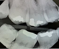
Figure 1 indicates that the PID is not parallel to the interproximal surfaces, causing severe horizontal overlap in both the maxilla and mandible. The radiographic image would be considered undiagnostic. The ideal method used for an excellent bitewing is to have the central beam parallel to proximal surfaces.
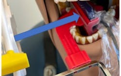
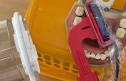
Literature suggests using the “open-door method” when contacts need to be clear and diagnostic.5 This method is achieved by moving the sensor distomesially by 15 degrees to ensure the canine is visible. This will keep the interproximal spaces open in the premolar bitewing. To avoid horizontal overlap, the central ray should be perpendicular to the arch of the teeth and directly through the interproximal spaces. With proper training and education on the concepts to mitigate overlapping, we can avoid exposing our patients to unnecessary radiation. Unfortunately, because the angle of the PID is off in Figure 1, the patient will need to be exposed to another radiograph. Proper angulation is shown in Figure 2, and proper positioning of the open-door method is shown in Figure 3.
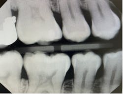
“Horizontal angulation refers to the positioning of the PID and the direction of the central ray in a horizontal, or side to side, plane.”6 Paralleling, bisecting, and bitewings all use similar principles of horizontal angulation and will not change according to technique. The PID needs be directed toward the center of the ring using a horizontal/parallel technique. With this correct angulation, proximal spaces will be cleared, and the radiopaque enamel with either be just touching or have a distant radiolucent line of separation. This will produce a diagnostic image (figure 4) where calculus or caries lesions will not go undetected.
If the PID is shifted too mesially or distally, horizontal overlap will occur. It is necessary to adjust the angulation of the tube head either in the mesial or distal direction to avoid horizontal overlaps and clear the contacts. This requires repositioning the direction in which the PID is facing by simply inclining the PID toward the mesial or distal aspect; there is no specific degree.
“If the lingual cusp appears mesial to the facial cusp, the tube head was angled too far in the mesial direction in relation to the interproximal contact. To correct this horizontal overlap, the tube head needs to be shifted horizontally in a distal direction. If the lingual cusp was distal to the facial cusp, then shift the tube head horizontally in the mesial direction to open the interproximal area of interest.”7 If vertical angulation is the cause of overlap, we can move the PID by 10 degrees pointing down.
According to the rule of SLOB (same lingual, opposite buccal), the object that appears lingually moves in the same direction of the PID. If the object appears in the opposite direction of the PID, the object would be on the buccal aspect. The rule of SLOB requires that two images of the same area be compared to determine the correct direction for the tube head. This will require a second radiograph or dental image.
On the second exposure, we modify the horizontal or vertical angulation to see where the object is located using the first image as a guide. For example, think of a dentist trying to locate lingual and buccal root canals.6 We would expose the patient twice, and then we can move mesially or distally just slightly in the second image. When the canal stays in the same direction whether mesial or distal, it would be known as the lingual canal, same lingual. The canal that moves in the opposite direction radiographically would be the buccal canal.
Summary
The goal and basic concepts of diagnostic horizontal bitewings are to reinforce ALARA.5,7 Proper utilization of radiographic techniques and clinician training will result in effective diagnostic horizontal bitewings. Instruments such as the Rinn XCP or the loop will assist in better placement. Continued practice, review, and accurate use of SLOB and the “open-door method” techniques will increase the clinician’s ability to take diagnostic bitewing radiographs or dental images and reduce unnecessary radiation. Occasionally it will be necessary to take a second x-ray if there is too much overlap or the radiograph is not diagnostic. This is not contrary to ALARA as radiographs need to be diagnostic.
Editor's note: This article appeared in the May 2023 print edition of RDH magazine. Dental hygienists in North America are eligible for a complimentary print subscription. Sign up here.
References
- Takahashi N, Lee C, Da Silva JD, et al. A comparison of diagnosis of early stage interproximal caries with bitewing radiographs and periapical images using consensus reference. Dentomaxillofac Radiol. 2019;48(2):20170450. doi:1259/dmfr.20170450
- Healthy People 2030. Oral conditions: overview and objectives. U.S. Department of Health and Human Services. Office of Disease Prevention and Health Promotion. Accessed July 6, 2022. https://health.gov/healthypeople/objectives-and-data/browse-objectives/oral-conditions
- Rasid L, Razak S, Ayoub AA, Kamaruzaman M, Azmi NW. Evaluation of image quality of bitewing radiographs taken by UiTM dental students. Compendium of Oral Science. 2020;7:32-41. doi:24191/cos.v7i0.17492
- Kositbowornchai S, Phadannorg T, Permpoonsinsook M, Thinkhamrop B. Bitewing film quality: a clinical comparison of the loop vs. holder techniques. Quintessence Int. 2004;35(4):321-325.
- Ostrander SO. Take it right the first time. RDH magazine. August 1, 2018. Accessed January 6, 2022. https://www.rdhmag.com/patient-care/article/16408259/take-it-right-the-first-time
- Iannucci JM, Howerton LJ. Dental Radiography Principles and Techniques. 5th ed. Saunders; 2016.
- Mauriello SM. Prevent technique errors. Dimens Dent Hyg. June 10, 2016. Accessed January 6, 2023. https://dimensionsofdentalhygiene.com/article/prevent-technique-errors/


