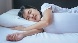
Bruxism is often seen in the dental office and is listed as the “third most common form of sleep disorders after sleep talking and snoring.”1 This parafunctional habit (or parasomnia in medical terms) of grinding or gnashing the teeth and clenching the jaw has two different subdisorders—awake (diurnal) bruxism and sleep (nocturnal) bruxism—and is encompassed by a complex web of supposed causes and variables. Sleep bruxism (SB) exists in 8% to 31.4% of the population, while awake bruxism has a higher prevalence exhibited in 22.1% to 31% of the general population.2
Unfortunately, both conditions have the same deleterious effects on the patient’s mouth and jaw, causing a cascade of destructive symptoms in the mouth, head, and neck. The parafunctional activities of bruxism cause hypersensitivity in teeth, headaches, painful muscles of the jaw and temporomandibular joint (TMJ), occlusal wear, and often, damage dental restorations, even dental implants. Indeed, 13% of failed implants are attributed to bruxism, making recognition of the disorder essential before commencing implantation work.3
Without question, bruxism is a constant symptom in the dental office, at least in its presenting symptoms. However, there is more to this complex and perplexing disorder than meets the eye, as any dental professional who has been in the field for more than a few years can tell you. Beyond the local effects, the syndrome is correlated with a host of other medical and lifestyle issues. This leads us to the question: Is bruxism a medical or dental issue?
The signs
The first signs you notice in your patient’s mouth are occlusal wear and thinning of the enamel, which can expose the sensitive underlying dentin and alter the patient’s vertical bite dimensions. Sometimes this can be from historical bruxing, and the patient may no longer have this habit. Using the Parafunctional Risk Rating system (PRR) can help to explore this further (table 1).4 Often, patients themselves may not even be aware of the bruxing unless a family member or partner happens to awaken them and hear the alarming popping and grinding sounds coming from the mouth.
The exact prevalence of SB is hard to determine as most bruxers (more than 80%) are unaware of their habit. Over time, wear patterns become more prominent with a noticeable flattening of the teeth, especially the canines, incisors, and even back to the molars.5 Each bruxism episode consists of repeated rhythmic masticatory muscle activity (RMMA) repeated rapidly, applying a powerful force on the enamel. Eventually, you will discover a barrage of damage, such as tooth chips, breaks, fractures, mandibular and maxillary tori, increased tooth mobility, increased muscle tone in the jaw, morning muscle fatigue for sleep bruxers, and TMJ discomfort.6 These are all indicators of bruxism and its consequences, but what causes the onset of this condition?
Contributors to bruxism
Multiple causes of bruxism have been hypothesized and studied, but there are still many unanswered questions. Initially, the condition was theorized to be due to uneven dental occlusion, but it was later categorized as a sleep disorder. Dentists would consider it an airway issue, but medical science doesn’t wholly agree. The fact that two different systems are at play—respiratory and neurological—makes it more complicated.
In reality, bruxism can be attributed to a wide variety and combination of factors, but there has been no conclusive testing that reveals a primary and consistent cause. Medically speaking, there appears to be a relationship between bruxing and neurological disorders, including migraine, headaches, and multiple sclerosis.7 Gastroesophageal reflux disease (GERD) is another contributing factor, and studies are increasingly showing a link between hypersomnia (excessive sleepiness during the day) and cardiovascular issues.8
The issue of bruxism may sometimes arise as a result of a traumatic brain injury or as part of a neurological disorder, such as Parkinson’s disease, dementia, or Alzheimer’s disease. Psychosocial issues, such as stress, anxiety, anger, and tension, are one group of potent influences. Awake bruxism, specifically, is associated with the stress caused by family responsibilities or work pressures.9 It is estimated that “nearly 70% of bruxism cases overall are the result of stress or anxiety.”10 Personality types with aggressive, controlling, or competitive behaviors also show increased bruxism.11 In combination with these mental and emotional factors, certain antidepressants known as selective serotonin reuptake inhibitors (SSRIs), such as fluoxetine and paroxetine, can cause bruxism in patients of any age, including children.12 A patient’s mental state heavily influences rates of bruxism as do other lifestyle choices, such as smoking tobacco, drinking alcohol,13 or using recreational drugs, such as MDMA.12 During dental assessments, professionals need to investigate their clients’ mental states, daily habits, and any prescriptions they are currently taking. While a patient’s genetic makeup is out of his or her control, it is another possible contributor to the development of bruxism.14
Nocturnal bruxism is currently classified as a sleep-related movement disorder.16 Sleep disorders and sleep arousal cycles show a strong link with SB in recent studies.17 Research shows that sleep apnea and SB tend to co-occur, but researchers have not yet been able to produce conclusive evidence to understand the actual relationship between sleep apnea and SB.18
Bruxism and sleep arousal patterns, in which a person shifts suddenly to another lighter REM state or even wakes up, have a connection as well.19 Researchers have discovered that SB sufferers show higher bruxism episodes during the ascending phase of the sleep cycle.20 This sleep disturbance can add to anxiety and low mood as well as cause a vicious cycle of stress and bruxism. Even vitamin D deficiency has been linked to this movement and general sleep disorder.21 Although sleeping supine is a favored position for many people, this sleep position can also increase the likelihood of bruxism.22 Another theory suggests neurotransmitters and the central nervous system are responsible for the ailment, with evidence that various neurotransmitters modulate SB in the central nervous system, and they can malfunction.10 Specifically, the basal ganglia are suggested to be responsible for hyperactivity during nocturnal bruxism, which has to do with the theory of neuroplasticity.23
Course of action treatment plans
Many of these influences are outside the dental realm, but as professionals, it is up to us to study the research and complete a proper analysis so we can counsel our patients on how to best manage this disorder. It may be time for us to take a more holistic approach and consider the factors causing bruxism rather than just prescribing a mouthguard to prevent damage. Historically, the dental community has been perplexed, using many different methodologies to treat nocturnal and diurnal bruxism. Some dentists merely address and repair the symptoms, whether they are broken teeth, abfraction lesions, or sensitive teeth, and ignore the underlying root causes. Other practitioners are more proactive and utilize standards such as the PRR4 to identify a patient’s risk level and develop a treatment strategy to avoid extreme occlusal wear, enamel damage, painful TMJ, and muscular issues.
Current treatments range from counseling for lifestyle changes (e.g., alleviating stress, avoiding alcohol and tobacco, or changing antidepressants) to more physically focused approaches, such as occlusal adjustments, custom, stock, or boil-and-bite mouth guards, pharmaceutical therapies, and even Botox. Biofeedback therapy has shown some promising results, with scientists developing a miniature self-contained EMG detector with a biofeedback function for the nocturnal variant.24 Although this is not in mainstream use yet, it is a possible option for the future. Dentists addressing restorations must pay attention to their patient’s level of bruxing as porcelain often breaks down. Some utilize metal-based substructures instead, to protect from microcracks.25
With so many options out there to address the problem, it can be confusing to choose the best course of treatment for your patient. However, if your dental team can come together during your patient’s chair time and arrive at a comprehensive and complete understanding of the situation through occlusal analysis, evaluation of TMJ and the masticatory muscles, and investigation of the patient’s lifestyle, habits, and symptoms, it is possible to produce a plan that can change the patient’s life for the better. Providing therapies that improve our patients’ lives should be the highest goal of our vocation.
The future of bruxism treatment
Unequivocally, bruxism is both a medical and a dental issue. This affliction spans a wide array of possible variants, weaving in genetics, lifestyle, pharmaceutical usage, personality, vitamin deficiency, sleep cycles, and neurological threads. Bruxism has proven itself to be a complex parafunctional syndrome that needs further research to narrow down its primary causes and most effective treatment options. This will likely take years of further scientific study.
What our patients need now is a proactive and holistic approach to assist them until the necessary research provides all the answers. If we operate as a team in the office and the wider oral community to manage bruxism and its detrimental parafunctional habits, we can bring hope for the management of this disorder.
It is time we dug down into the underlying causes of this mysterious parafunctional syndrome to improve our treatment recommendations and reach a consensus of methodologies in the dental community. During those busy, overwhelming days, there can be a tendency to rush our patients through their appointments and focus solely on their immediate physical treatment. As practitioners, we must learn to look holistically at the entire body and other pathophysiology that co-occurs with dental issues, especially in the case of bruxism. Reductionist tendencies in the medical field can get us stuck in one corner of the body while ignoring other significant factors that are causative elements.
The dental industry and other medical fields are moving toward a more integrative style of practice. Our patients want more in-depth care with knowledgeable insight into the interplay of many components of the body and its correlated mental states. A mind-body complex is rising to the foreground of our world. As professionals, if we do not want to be left behind, we need to expand our view of dentistry and therapy in the office. It is our duty to understand our patients, empowering them to understand their oral health and help them to heal.
References
1. Murali RV, Rangarajan P, Mounissamy A. Bruxism: Conceptual discussion and review. J Pharm Bioallied Sci. 2015;7(suppl 1):S265-S270.
2. Manfredini D, Winocur E, Guarda-Nardini L, Paesani D, Lobbezoo F. Epidemiology of bruxism in adults: a systematic review of the literature. J Orofac Pain. 2013;27(2):99-110. doi:10.11607/jop.921
3. Kang DW, Kim SH, Choi YH, Kim YK. Repeated failure of implants at the same site: a retrospective clinical study. Maxillofac Plast Reconstr Surg. 2019;41(1):27. doi:10.1186/s40902-019-0209-1
4. Parafunction Risk Rating Protocol. Orofacial Therapeutics website. https://quicksplint.com/pages/parafunction-risk-rating-protocol. Accessed January 7, 2020.
5. Wall RT. The clues behind bruxism. RDH magazine website. https://www.rdhmag.com/pathology/temporomandibular-joint-dysfunction/article/16403831/the-clues-behind-bruxism. Published September 1, 2004. Accessed January 6, 2020.
6. Machado NA, Fonseca RB, Branco CA, Barbosa GA, Fernandes Neto AJ, Soares CJ. Dental wear caused by association between bruxism and gastroesophageal reflux disease: a rehabilitation report. J Appl Oral Sci. 2007;15(4):327–333. doi:10.1590/s1678-77572007000400016
7. Williams DE, Lynch JE, Doshi V, Singh GD, Hargens AR. Bruxism and temporal bone hypermobility in patients with multiple sclerosis. Cranio. 2011;29(3):178-186. doi:10.1179/crn.2011.026
8. Martynowicz H, Dymczyk P, Dominiak M, et al. Evaluation of intensity of sleep bruxism in arterial hypertension. J Clin Med. 2018;7(10). doi:10.3390/jcm7100327.
9. Manfredini D, Lobbezoo F. Role of psychosocial factors in the etiology of bruxism. J Orofac Pain. 2009;23(2):153-166.
10. Arakelyan HS. Neurotransmitters, sleep bruxism, food and drugs. https://www.researchgate.net/publication/320064956_Neurotransmitters_Sleep_BruxismFood_and_Drugs. Published September 27, 2017. Accessed January 6, 2020.
11. Wieckiewicz M, Paradowska-Stolarz A, Wieckiewicz W. Psychosocial aspects of bruxism: The most paramount factor influencing teeth grinding. Biomed Res Int. 2014:469187. doi:10.1155/2014/469187. https://www.hindawi.com/journals/bmri/2014/469187/. Published July 13, 2014. Accessed January 7, 2020.
12. Garrett AR, Hawley JS. SSRI-associated bruxism: A systematic review of published case reports. Neurol Clin Pract. 2018;8(2):135-141. doi:10.1212/CPJ.0000000000000433
13. Bertazzo-Silveira E, Kruger CM, Porto De Toledo I, et al. Association between sleep bruxism and alcohol, caffeine, tobacco, and drug abuse: A systematic review. J Am Dent Assoc. 2016;147(11):859-866. doi:10.1016/j.adaj.2016.06.014
14. Dinis-Oliveira R, Caldas I, Carvalho F, Magalhães T. Bruxism after 3,4-methylenedioxymethamphetamine (ecstasy) abuse. Clin Toxicol. 2010;48(8):863-864. doi:10.3109/15563650.2010.489903
15. Lobbezoo F, Visscher CM, Ahlberg J, Manfredini D. Bruxism and genetics: A review of the literature. J Oral Rehabil. 2014;41(9):709-714. doi:10.1111/joor.12177
16. Carra MC, Huynh N, Lavigne G. Sleep bruxism: a comprehensive overview for the dental clinician interested in sleep medicine. Dent Clin North Am. 2012;56(2):387-413. doi:10.1016/j.cden.2012.01.003
17. Ohayon NM, Li KK, Guilleminault C. Risk factors for sleep bruxism in the general population. Chest. 2001;119(1):53-61. doi:10.1378/chest.119.1.53
18. Winck M, Drummond PM, Viana P, Pinho JC, Winck JC. Sleep bruxism associated with obstructive sleep apnoea syndrome – A pilot study using a new portable device. Rev Port Pneumol. 2017;23(1):22-26. doi:10.1016/j.rppnen.2016.07.001
19. Lavigne GJ, Khoury S, Abe S, Yamaguchi T, Raphael K. Bruxism physiology and pathology: an overview for clinicians. J Oral Rehabil. 2008;35(7):476-494. doi:10.1111/j.1365-2842.2008.01881.x
20. Huynh N, Kato T, Rompré PH, et al. Sleep bruxism is associated to micro-arousals and an increase in cardiac sympathetic activity. J Sleep Res. 2006;15:339-346. doi:10.1111/j.1365-2869.2006.00536.x
21. Gominak SC, Stumpf WE. The world epidemic of sleep disorders is linked to vitamin D deficiency. Med Hypotheses. 2012;79(2):132-135. doi:10.1016/j.mehy.2012.03.031.
22. Miyawaki S, Lavigne GJ, Pierre M, Guitard F, Montplaisir J, Kato T. Association between sleep bruxism, swallowing-related laryngeal movement, and sleep positions. Sleep. 2003;26(4):461-465.
23. Behr M, Hahnel S, Faltermeier A, et al. The two main theories on dental bruxism. Ann Anat. 2012;194(2):216-219. doi:10.1016/j.aanat.2011.09.002
24. Kanathila H, Pangi A, Poojary B, Doddamani M. Diagnosis and treatment of bruxism: Concepts from past to present. Int J Appl Sci. 2018;4(1):290-295. http://www.oraljournal.com/pdf/2018/vol4issue1/PartE/4-1-44-680.pdf. Accessed January 7, 2020.
25. Raigrodski AJ. Cosmetic dentistry: The full mouth fixed rehabilitation of the bruxing patient—achieving function and esthetics. OralHealth website. https://www.oralhealthgroup.com/features/cosmetic-dentistry-the-full-mouth-fixed-rehabilitation-of-the-bruxing-patient-achieving-function-an/. Published April 1, 2001. Accessed January 7, 2020.


