Authors: Michele White, DDS, Bridget B. Huynh, Amber B. Hua, Royce Strickland, DDS, Cleverick (C.D.) Johnson, DDS, MS, Ben F. Warner, DDS, MD, MS
The new-patient dental exam, or complete oral exam, is an important aspect of the establishment of the relationship between the patient and the dental team. During this initial visit, it is the duty of dental personnel to gather information for the safe and effective treatment of the patient.
Along with the medical and dental history, extraoral and intraoral clinical exams are performed. Observation of clinical findings—and discerning whether they are within normal limits or atypical—launches the treatment planning phase. The fingernails, as a readily observable feature, can aid the dental hygienist and dentist when considering the patient’s overall health.
Do you really know what oral-systemic means?
Eye spy: Exploring the mouth-eye connection
A literature review of commonly found diseases that are clinically relevant and observed in the nails was conducted for this study. Dental professionals can observe features in the nails of their patients as an aid to assist them in differentiating their patients’ health. Following are examples of commonly found diseases that have presentation in the fingernails.
Mees’ lines, Beau’s lines, Muehrcke’s lines
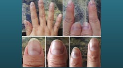
There are several atypical presentations of the nails that can suggest disease to the dental team; e.g., Beau’s lines, Mees’ lines, and Muehrcke’s lines (figures 1-3).1 Although they all present in a transverse horizontal manifestation, differences can be seen among these abnormalities.
Beau’s lines exhibit depressions or grooves that are the result of conditions that cause the nail to slow or stop growing. Both Mees’ lines and Muehrcke’s lines present as a white lesion in the nail bed.2 Only Beau’s lines and Mees’ lines move distally with nail growth.3 Beau’s lines, Mees’ lines, and Muehrcke’s lines are associated with systemic illness, trauma, poisoning, medications, and high-altitude mountaineering.2
COVID nails
With the onset of the SARS-CoV-2 (COVID-19) pandemic, mucocutaneous expression of illness presented and is still being documented.4 One such symptom presents in the nail beds and is referred to as COVID nail or COVID red half-moon nail (figure 4).4 COVID nail displays as a half-moon-shaped transverse red/violet band above the nail lunule5 with an onset as early as two days after COVID symptoms begin and as late as one month.6 Symptoms usually disappear once the patient has recovered from COVID-19.
Green nail syndrome
Another symptom of infection, green nail syndrome, occurs in the nail beds. It is seen most commonly as an occupational disease in health-care workers, especially in intensive care units, and lately, during the COVID-19 pandemic.7,8 Treatment includes topical and/or systemic antibiotics, diluted sodium hypochlorite, antifungals, and tobramycin eye drops.7
Koilonychia
Koilonychia is a convex, spoon-shaped aberration of the nail that is most often caused by iron-deficiency anemia (figure 5).9 Though considered the world’s most common nutritional deficiency disease, this symptom is often secondary to lack of folate, protein, or vitamin C. Other possible causes include autoimmune diseases, such as celiac disease, that cause an inability to absorb iron from food; hypothyroidism; low blood supply to the extremities; vitamin B deficiency; and cancer.10
Digital hypertrophic osteoarthropathy or digital clubbing
Digital hypertrophic osteoarthropathy (HOA), or clubbing of the fingers, presents as an enlargement of the ends of the fingers (figure 6). It commonly occurs bilaterally as a primary or secondary condition. Primary HOA, or pachydermoperiostosis, is a hereditary condition.11 Secondary HOA is associated with disease, including lung, cardiovascular, inflammatory, cancer, thyroid, and liver diseases.12 The most common finding in children is HOA secondary to cystic fibrosis.12
Terry’s nails
Terry’s nails present with a whitish or “ground glass” appearance, an absence of the lunula, and a normal or sometimes reddish-brown discoloration at the distal end of the nail (figure 7).
This symptom is often secondary to cirrhosis, chronic renal failure, and congestive heart failure. There is no treatment as symptoms will decrease when the underlying condition improves.13
Yellow nail syndrome
Yellow nail syndrome (YNS) is a rare disorder that is associated with the pulmonary manifestations of chronic cough, bronchiectasia, and pleural effusion. Lower limb lymphedema due to lymphatic impairment and chronic sinusitis are also common causes. YNS can resolve on its own and/or with the aid of medicaments such as antifungals and oral vitamin E.14
Cyanosis
Central cyanosis of the nails is secondary to poor arterial oxygenation.15 Appearing also in the skin, tongue, and mucous membranes, this symptom should alert the dental team to further understand the patient’s medical history (figure 8).
Conclusion
The nail beds, as a readily observable clinical feature, can present according to a person’s health at the time of the dental exam. Understanding differences among the clinical presentations can assist the dental hygienist and dentist in recognizing variations from normal that can support the comprehensive oral exam. Additionally, having a baseline medical history, dental history, extraoral and intraoral findings, hard- and soft-tissue findings, including the nail beds, can assist the dental team in observing changes in the clinical presentation of the patient’s oral and medical health. Especially useful at the initial visit, observation of the nail beds can serve both the patient and the dental team.
Editor's note: This article appeared in the December 2022 print edition of RDH magazine. Dental hygienists in North America are eligible for a complimentary print subscription. Sign up here.
References
- Sharma V, Kumar V. Muehrcke lines. Can Med Assoc J. 2013;185(5):E239. doi:10.1503/cmaj.120269
- Jandial A, Mishra K, Prakash G, Malhotra P. Beau’s lines. BMJ Case Rep. 2018;2018:bcr2018224978. doi:10.1136/bcr-2018-224978
- Aujayeb A. Mees’ lines in high altitude mountaineering. BMJ Case Rep. 2019;12(3):e229644. doi:10.1136/bcr-2019-229644
- Gaurav V, Grover C. “COVID” terminology in dermatology. Indian J Dermatol. 2021;66(6):707. doi:10.4103/ijd.IJD_472_21
- Méndez-Flores S, Zaladonis A, Valdes-Rodriguez R. COVID-19 and nail manifestation: be on the lookout for the red half-moon nail sign. Int J Dermatol. 2020;59(11):1414. doi:10.1111/ijd.15167
- Thakur V, Bisht YS, Sethi S, Jindal R. Red nail bands in conjunction with telogen effluvium as a post-COVID-19 phenomenon. Australas J Dermatol. 2022;63(1):141-142. doi:10.1111/ajd.13779
- Heymann WR. Going green: the complexities of the green nail syndrome. Dermatol World Insights Inquiries. 2021;3(5).
- Wollina U, Kanitakis J, Baran R. Nails and COVID-19—a comprehensive review of clinical findings and treatment. Dermatol Ther. 2021;34(5):e15100. doi:10.1111/dth.15100
- Ghaffari S, Pourafkari L. Koilonychia in iron-deficiency anemia. N Engl J Med. 2018;379(9):e13. doi:10.1056/NEJMicm1802104
- Wilson DR, Metropulos M. Koilonychia: why are my nails spoon shaped? Medical News Today. November 19, 2019. https://www.medicalnewstoday.com/articles/318761
- Leader D, Jelic S. Clubbing of the fingers or toes. VeryWell Health. July 24, 2022. https://www.verywellhealth.com/clubbing-of-fingers-914776
- Essouma M, Nkeck JR, Agbor VN, Noubiap JJ. Epidemiology of digital clubbing and hypertrophic osteoarthropathy–a systematic review and meta-analysis. J Clin Rheumatol. 2022;28(2):104-110. doi:10.1097/RHU.0000000000001830
- Witkowska AB, Jasterzbski TJ, Schwartz RA. Indian J Dermatol. Terry’s nails: a sign of systemic disease. 2017;62(3):309-311. doi:10.4103/ijd.IJD_98_17
- Vignes S, Baran R. Yellow nail syndrome: a review. Orphanet J Rare Dis. 2017;12(1):42. doi:10.1186/s13023-017-0594-4
- Changizi M, Rio K. Harnessing color vision for visual oximetry in central cyanosis. Med Hypotheses. 2010;74(1):87-91. doi:10.1016/j.mehy.2009.07.045
- Beau’s lines on left middle fingernail. Wikimedia Commons. January 12, 2014. Accessed November 14, 2022. https://commons.wikimedia.org/wiki/File:Beau%27s_line_on_left,_middle_fingernail.jpg
- Trottier Y. Mees’ lines. Wikimedia Commons. July 14, 2012. Accessed November 14, 2022. https://commons.wikimedia.org/wiki/File:Mees%27_lines.jpg
- Chemotherapy-induced Muehrcke’s lines on a 28-year-old female. Wikimedia Commons. July 2009. Accessed November 14, 2022. https://commons.wikimedia.org/wiki/File:Muehrcke%27s_lines.JPG
- CHeitz. Iron deficiency anemia. Flickr. April 14, 2014. https://www.flickr.com/photos/coreyheitzmd/15023020192/in/photolist
- Desherinka. Clubbed fingers. Wikimedia Commons. August 18, 2019. Accessed November 14, 2022. https://commons.wikimedia.org/wiki/File:Dedos_con_acropaquia.jpg
- Hojasmuertas. Terry’s nails. Wikimedia Commons. October 2, 2012. Accessed November 14, 2022. https://commons.wikimedia.org/wiki/File:Terry%27s_nails.jpg
- Heilman J. Cyanosis of the hand in someone with low oxygen saturations. Wikimedia Commons. October 23, 2011. https://commons.wikimedia.org/wiki/File:Cynosis.JPG


