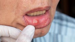
Recurrent aphthous stomatitis (RAS), often known as canker sores, is a common oral condition characterized by the repeated appearance of painful ulcers. These lesions can significantly impact quality of life; they typically present in small, round, or oval-shaped ulcers with well-defined erythematous edges that tend to occur in clusters, covered with a yellow/gray fibrinous pseudomembrane.1
They most commonly affect the buccal mucosa, tongue, lips, and floor of the mouth. They are often accompanied by a burning/tingling sensation that may precede the ulcers, pain (particularly when eating or speaking), and discomfort while eating spicy and acidic foods.2
Gluten sensitivity (GS) and canker sores: A dance of inflammation
Over the past 30 years, increased awareness and improved diagnostic tools have led to a surge in celiac disease diagnoses. Despite this advancement, a vast majority—estimated to be 95%—remain undiagnosed.3
Gluten is naturally in grains such wheat, rye, and barley. It plays a key role in food structure, but people react differently to it. With celiac disease, the most severe form of gluten sensitivity, gluten triggers an autoimmune response, prompting the body to attack its own tissues. This assault isn't confined solely to the gut; it can extend beyond, potentially impacting the oral mucosa and setting the stage for aphthous stomatitis.
While the full relationship between gluten and canker sores remains a puzzle, several theories offer clues. Autoimmunity could be a prime suspect. Celiac disease's inflammatory response might spill over to the mouth, targeting the epithelial cell lining and disrupting its protective barrier.4 This weakened defense leaves the mucosa vulnerable to irritation and the formation of painful ulcers.
Another potential culprit is nutrient deficiency. GS can hinder nutrient absorption, leading to deficits in iron, folate, and B vitamins. These deficiencies weaken the oral mucosa, compromising its protective capacity and paving the way for these lesions.
A tangible link?
There could be more subtle factors beyond these direct mechanisms. Inflammatory ripples triggered by gluten, even in nonceliac sensitivity, could contribute to a generalized low-grade inflammation affecting the entire body, including the mouth.
The gut-brain axis also enters the plot. Our gut health directly influences our neurological well-being, and emerging research suggests that conditions such as gluten sensitivity can impact the nervous system, including the trigeminal nerve responsible for facial sensations such as pain. This altered nerve signaling might play a role in the formation and persistence of mouth sores.5
Regardless of their tie to gluten, canker sores are common and have numerous other causes. Stress, vitamin deficiencies,6 trauma, and certain medications can all contribute to these painful lesions.
Despite the current unknowns, some things are clear. Canker sores are significantly more prevalent among people with celiac disease, suggesting a tangible link. For some people with GS, adopting a gluten-free diet can alleviate or even resolve the lesions, implying a dietary component to the puzzle.7
Our role as dental professionals is to have a keen eye and make a comprehensive assessment that can pave the way for further investigations, such as blood tests for celiac-specific antibodies or endoscopy for intestinal tissue evaluation. This collaborative approach between the dental community and gastroenterologists is vital in unraveling the complex web of factors contributing to RAS and ultimately delivering accurate diagnoses and effective patient treatment strategies.
References
1. Rodrigo L, Beteta-Gorriti V, Alvarez N, Gómez de Castro C, de Dios A, Palacios L, Santos-Juanes J. Cutaneous and mucosal manifestations associated with celiac disease. Nutrients. 2018;10(7):800. doi:10.3390/nu10070800
2. Giannetti L, Generali L, Bertoldi C. Oral pemphigus. G Ital Dermatol Venereol. 2018;153(3):383-388. doi:10.23736/S0392-0488.18.05887-X
3. Sahin Y. Celiac disease in children: A review of the literature. World J Clin Pediatr. 2021;10(4):53-71. doi:10.5409/wjcp.v10.i4.53
4. Milia E, Sotgiu MA, Spano G, Filigheddu E, Gallusi G, Campanella V. Recurrent aphthous stomatitis (RAS): guideline for differential diagnosis and management. Eur J Paediatr Dent. 2022;23(1):73-78. doi:10.23804/ejpd.2022.23.01.14
5. Corriero A, Giglio M, Inchingolo F, Moschetta A, Varrassi G, Puntillo F. Gut microbiota modulation and its implications on neuropathic pain: a comprehensive literature review. Pain Ther. https://link.springer.com/article/10.1007/s40122-023-00565-3
6. Sardana K, Sachdeva S. Role of nutritional supplements in selected dermatological disorders: a review. J Cosmet Dermatol. 2022;21(1):85-98. doi: 10.1111/jocd.14436
7. Shakeri R, Zamani F, Sotoudehmanesh R, Amiri A, Mohamadnejad M, Davatchi F, Karakani AM, Malekzadeh R, Shahram F. Gluten sensitivity enteropathy in patients with recurrent aphthous stomatitis. BMC Gastroenterol. 2009;9:44. doi: 10.1186/1471-230X-9-44
Andreina Sucre, MSc, RDH, is an oral pathology and oral surgery specialist, and a practicing dental hygienist in South Florida. She’s a speaker, writer, and advocate for early pathological diagnosis. Andreina was born in Venezuela where she earned her dental degree in 2002 from Universidad Santa Maria. In December 2005, she earned her certificate and specialization degree in oral pathology and oral surgery from Pontificia Universidad Javeriana, in Colombia. Follow her on Instagram @ThePathoRDH.


