BY NANCY W. BURKHART, BSDH, EdD
Your patient today is Paul. He is 10 years old and has been a patient of record since age two. Paul has been diagnosed with regional odontodysplasia (RO). You will complete an updated medical history with the help of his parents and also perform a thorough intraoral and extraoral exam. The other hygienist in your office usually treats Paul, so you quickly try to remember what you learned about RO in college.
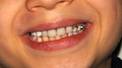
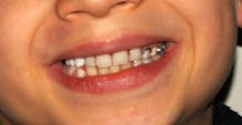
His parents report that Paul had been diagnosed at 10 months with rickets and also perinatal encephalopathy. He also had repeated infections up until the age of three and multiple tooth infections were noted in early life as well (see Figure 1).
The chief complaint from the parents is the continued failure of eruption with various teeth and also the cosmetic appearance of the teeth. But, Paul has done very well during treatment and willingly smiles, displaying his teeth.
His treatment has consisted of some extractions with the placement of a partial denture to aid in tooth replacement and bone development. During your extraoral exam and review of his prior radiographs, you notice radiolucent areas in the posterior region that contain some slightly calcified structure, all having a very abnormal appearance. The term “ghost-like teeth” comes to mind as you view the early radiographs of Paul and you begin to remember what you learned about regional odontodysplasia in college.
Regional odontodysplasia is considered a rare, severe developmental anomaly affecting the formation of the teeth. RO affects the structures derived from epithelial and mesenchymal components of the teeth, which include enamel, dentin, pulp, and the dental follicle. The roots are short with open apices and the pulp appears much larger than normal with little definition or contrast within the tooth structures.
Although the etiology of this entity is not completely known or understood, we do know that it is not hereditary. One theory is that the tooth or teeth within an adjacent area do not have the complete blood supply that is needed to calcify the structure; therefore, the enamel and dental matrix cannot be fully formed. Other reported etiology lists somatic mutations affecting the dental lamina; vascular disorders; pharmacological treatments during pregnancy; local or systemic viral infections; activation of latent viruses or differentiation failures of the neural crest cells; local trauma; nutritional deficiency; radiation; hyperpyrexia; local ischemia; and RH compatibility (Barberìa et al., 2012).
RO usually affects one arch and may be isolated to a particular tooth region that is innervated by certain nerves and blood vessels. Typically, tooth involvement does not cross the midline of the arch that is affected. A bimodal peak is documented usually at the times of eruption of the deciduous teeth or the permanent teeth. Most commonly observed with this entity are the maxillary anterior teeth. However, others have reported cases in the mandible, and the disorder is reported in both sexes, although the literature supports a slight female predilection. Studies by Barberìa et al. (2012)reported the appearance of RO in several quadrants of three male patients, affecting the mandible. Tooth involvement crossed the midline of the arch, and both deciduous and permanent teeth were affected.
When a large region is affected with more than one quadrant, the term generalized odontodysplasia is used. The affected teeth may appear rough, streaked, with deep grooves, and they may be poorly formed. The color may vary with yellow or brown hues and may appear hypoplastic or hypocalcified (Mehta DN et al., 2011). A differential diagnosis would likely include amelogenesis imperfecta, dental dysplasia, amelogenesis, as well as other disease states affecting the tooth formation apparatus.
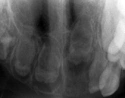
Because of the limited areas affected, diagnosis is usually made with regard to RO by clinical and radiographic analysis. Radiographically, the teeth are seen as poorly developed with very little dentin or enamel showing calcification, thereby presenting with a less radiopaque appearance than a normally developed tooth. Because of this poor development and the lack of calcification, they have been termed “ghost teeth” with only a shadow of a tooth present (see Figure 2).
Treatment and prognosis: Caries and inflammation are common in erupted teeth that are affected by RO and the incidence of a bacterial infection is increased in this patient group as well. Protecting the crown in erupted teeth is important for the long-term retention of the tooth. The prime consideration and treatment is directed toward retention of the teeth in order to encourage the growth of the alveolar ridge. The presence of teeth is important during the skeletal growth phase. Erupted teeth are treated very carefully so that as much calcification or hard tissue can be maintained and encouraged. However, extractions are often needed when salvaging the tooth is not possible. In some cases, the risk of infection and poor prognosis make extraction the only option.
With children especially, the psychological consequences of missing teeth or poorly formed teeth is a prime consideration. Other children often comment about physical appearances and the child may become withdrawn or feel different than his/her peers. Cosmetic procedures may be undertaken when possible. Partial dentures are made and must be changed or adjusted during the growth phase.
Ponranjini et al. point out that the best treatment options for these patients depends on the time of diagnosis, presenting symptoms, the functional and esthetic needs of each patient, and available treatment modalities. The hygienist may treat such patients and maintenance is very important in the long-term success of these patients with RO.
As always, keep asking good questions and always listen to your patients. RDH
References
Barberìa E, Coarasa AS, Hernandez A, Cardosa-Silva C. Regional odontodysplasia. A review of the literature and three case reports. Eur J Paediatr Dent. 2012; 13(2): 161-6.
Mehta DN, Bailoor D, Patel B. Regional odontodysplasia. J Indian Soc Pedod Prev Dent. 2011; 29:323-6.
Ponranjini VC, Jayachandran S, Bakyalakshmi K. Regional odontodysplasia: a report of a case. J Dent Child. 2012;79:26-9.
NANCY W. BURKHART, BSDH, EdD, is an adjunct associate professor in the department of periodontics, Baylor College of Dentistry and the Texas A&M Health Science Center, Dallas. Dr. Burkhart is founder and cohost of the International Oral Lichen Planus Support Group (http://bcdwp.web.tamhsc.edu/iolpdallas/) and coauthor of General and Oral Pathology for the Dental Hygienist. She was a 2006 Crest/ADHA award winner. She is a 2012 Mentor of Distinction through Philips Oral Healthcare and Pennwell Corp. Her website for seminars on mucosal diseases, oral cancer, and oral pathology topics is www.nancywburkhart.com.
Past RDH Issues


