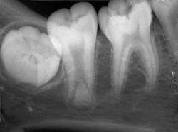
A 24-year-old male visited a dental office for an initial examination and routine checkup. Radiographic examination revealed an unusual mandibular second molar.
History
The patient denied any history of symptoms or pain associated with the mandibular molar area. The patient appeared to be in a general good state of health with no significant medical history. His dental history included sporadic checkups and routine restorative dental treatment.
Examinations
The patient's vital signs were all found to be within normal limits. Extraoral examination of the head and neck region revealed no enlarged or palpable lymph nodes. Intraoral examination revealed no abnormalities present.
Based on the clinical examination of the patient, selected periapical radiographs, bitewings, and a panoramic film were ordered and exposed. A review of the periapical films revealed a mandibular second molar with an elongated pulp chamber and an abnormally low furcation region (see radiograph). Clinically, the second molar appeared normal in size and shape. No other abnormalities were noted on the radiograph.
Clinical diagnosis
Based on the clinical and radiographic information available, which one of the following is the most likely diagnosis?
• dentin dysplasia
• odontoma
• taurodont
• macrodont
• regional odontodysplasia
Diagnosis
• taurodont
Discussion
The taurodont is a developmental anomaly that can be described as a tooth with an elongated, large pulp chamber and short roots with a low furcation area. The term taurodont refers to a tooth that is "bull-like" (tauro=bull, dont=tooth); the term is used to describe human teeth that resemble the molar teeth of cud-chewing animals. The exact cause of this abnormality is unknown. The taurodont may occur as an isolated trait or as a component of a syndrome. The taurodont may be associated with conditions such as amelogenesis imperfecta and ectodermal dysplasia, or seen with Klinefelter or Down's syndromes.
Clinical features
The taurodont is uncommon. The prevalence is estimated to be 2.5 percent to 3.2 percent of the U.S. population. There is no sex predilection. Although the taurodont may be seen in the deciduous dentition, the permanent dentition is affected more frequently. This anomaly typically affects a single molar tooth or multiple molars in the same quadrant and may occur unilaterally or bilaterally. The permanent second molar is most often affected. Clinically, the crown of the taurodont appears normal.
Radiographic features
A taurodont is recognized by its characteristic radiographic appearance. The overall tooth length is normal, but the distance from the cementoenamel junction to the furcation is increased. The pulp chamber is greatly enlarged without a constriction at the cementoenamel junction. The rectangular shape of the pulp chamber resembles that of cud-chewing animals. The roots appear to extend only a short distance from the furcation and exhibit short root canals. Radiographically, the taurodont tends to have an overall "stretched" appearance.
iagnosis and treatment
The diagnosis of a taurodont is made based on its radiographic appearance. The radiographic appearance of a taurodont may be confused with that of an enlarged pulp chamber seen in association with a developing tooth; the open apices of the developing tooth should enable the practitioner to distinguish between the two. The taurodont does not require treatment; however, conditions and syndromes that are associated with taurodonts should be ruled out. The enlarged pulp chamber can be a complicating factor during root canal procedures.
Joen Iannucci Haring, DDS, MS, is a professor of clinical dentistry, Section of Primary Care, The Ohio State University College of Dentistry.


