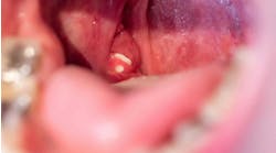
initial examination. Oral examination revealed a generalized brown-colored dentition.
Joen Iannucci Haring, DDS, MS
History
The patient stated that his teeth had appeared brown for as long as he could remember. When questioned if other family members had similar-appearing teeth, he stated that his father also has brown-colored teeth.
The patient appeared to be in an overall good state of health at the time of the dental visit. No significant problems were noted during the medical history and no medications were being taken by the patient at the time of the dental examination.
Examinations
No unusual or abnormal findings were identified during the extraoral examination. Intraoral examination revealed a generalized brown-colored dentition (see photo). Examination of the oral soft tissues revealed no unusual findings and no bony abnormalities. Radiographic examination revealed teeth with thin amounts of enamel and areas where the enamel had fractured away (see radiograph).
Clinical diagnosis
Based on the information provided, which one of the following is the most likely diagnosis?
* Regional odontodysplasia
* Dentin dysplasia
* Amelogenesis imperfecta
* Dentinogenesis imperfecta
* Internal resorption
Diagnosis
Amelogenesis imperfecta
Discussion
Amelogenesis imperfecta is an inherited disorder characterized by abnormal enamel formation. The term amelo refers to enamel and genesis means formation. Amelogenesis imperfecta refers to the imperfect formation of enamel. Amelogenesis imperfecta affects only the enamel; all other components of the tooth are normal. Amelogenesis imperfecta affects both the primary and permanent dentitions.
Enamel is formed in three stages: (1) the enamel matrix is formed, (2) the enamel matrix is mineralized, and (3) enamel maturation or secondary mineralization occurs. Problems resulting in enamel defects may arise during any one of these stages. The major types of amelogenesis imperfecta correlate with the defects that occur in these stages.
The forms of amelogenesis imperfecta include the following: (1) hypoplastic, (2) hypomaturation, (3) hypocalcification, and (4) hypomaturation/hypoplastic.
Within these categories, there are 14 recognized subtypes of amelogenesis imperfecta based on the clinical, radiographic, histologic, and genetic features.
Clinical and radiographic features
The hypoplastic form is the most common type of amelogenesis imperfecta. In this form, there is a deficient amount of enamel. Depending on the subtype classification, the crowns of the teeth may appear as one of the following: opaque white, translucent brown, yellow-brown, pitted, or grooved. Radiographically, the teeth exhibit abnormally thin amounts of enamel, or completely lack enamel. The radiodensity of the thin enamel present is greater than that of dentin. Because of the lack of enamel, the teeth clinically exhibit excessive interdental spacing. The teeth often are described as resembling crown preparations.
In the hypocalcified form of amelogenesis imperfecta, the teeth are formed with a very soft enamel due to the incomplete mineralization of the enamel structure. A sufficient amount of enamel is present, but there is no significant calcification. In this type, the enamel is quickly lost shortly after the teeth erupt into the oral cavity.
Consequently, the underlying dentin becomes exposed and stains easily. The teeth affected by hypocalcified amelogenesis imperfecta appear honey-brown in color with a roughened-surface texture. Radiographically, the teeth appear to lack enamel, or the enamel is less radiodense than dentin.
In the hypomaturation form of amelogenesis imperfecta, the normal amount of enamel is present and some mineralization does occur. However, there is a decreased amount of secondary mineralization (maturation) that occurs in the enamel. As a result, the enamel is softer than normal and tends to chip away from the underlying dentin. Fracturing of the enamel is common.
The use of a dental explorer with pressure may pit the surface. In this type of amelogenesis imperfecta, the crowns contact interproximally. The affected teeth may appear chalky white and rough or grooved. Radiographically, the affected enamel exhibits a radiodensity that is similar to dentin.
In the hypomaturation/hypoplastic form of amelogenesis imperfecta there is both the lack of enamel and the lack of enamel maturation. The most significant defect is that of the lack of enamel. The thin enamel that is present exhibits a lack of secondary mineralization. The teeth appear yellowish with opaque mottling, pitting, and attrition.
Radiographically, the enamel and dentin appear to have a similar density. Large pulp chambers also may be seen.
Treatment and prognosis
The treatment and prognosis for teeth with amelogenesis imperfecta vary with the severity of the enamel involvement. Variants that include rapid enamel loss and attrition require full-coverage crowns as soon as it is practical. When excessive crown loss occurs, restoration is not possible.
In patients without sufficient crown length for restorations, extractions and fabrication of complete dentures may be the only alternative. In the types of amelogenesis imperfecta that demonstrate less enamel loss, the aesthetic appearance is often the primary concern. Full-coverage crowns can be used to improve the clinical appearance of the crowns.
Joen Iannucci Haring, DDS, MS, is an associate professor of clinical dentistry, Section of Primary Care, The Ohio State University College of Dentistry.


