By Joen Iannucci Haring
A 43-year-old male visited a dentist for an initial examination and checkup. An abnormally large and misshapen mandibular tooth was noted during the clinical exam.
History
The patient was aware of the large and misshapen tooth. He had been seen previously by another dentist for checkups and routine restorative treatment, and the dentist had commented on the unusual appearance of the large mandibular tooth.
The patient appeared to be in an overall good state of health at the time of the dental visit. No significant problems were noted during the medical history and no medications were being taken by the patient at the time of the dental examination.
Examinations
No unusual or abnormal findings were identified during the extraoral examination.
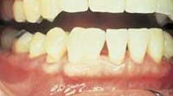
Intraoral examination revealed a large, misshapen tooth in the mandibular anterior region (see photo). Further examination revealed no other teeth in the dentition with a similar appearance. The patient's dentition consisted of a total of 27 teeth; 14 in the maxillary arch and 13 in the mandibular arch.
Examination of the oral soft tissues revealed no unusual findings, and no bony abnormalities were noted.
Clinical diagnosis
Based on the clinical information presented, which of the following is the most likely clinical diagnosis?
• gemination
• dilaceration
• macrodont
• concresence
• fusion
Diagnosis
• fusion
Discussion
The term fusion can be defined as "the abnormal joining of adjacent parts." Fusion is a developmental anomaly that results in the union of two adjacent tooth germs with confluence of dentin. True fusion always involves the union of dentin. The cause of fusion is uncertain; crowding, external pressure, and trauma have all been suggested.
Fusion may be complete or incomplete, depending on the stage of development of adjacent tooth germs at the time of contact. Early contact of adjacent tooth buds may result in a single, large tooth; later contact may result in the union of crowns or roots only.
Fusion occurs more frequently in the deciduous dentition than in the permanent dentition. Fusion most often involves the anterior teeth; the incisors are most often affected.
Clinical features
Clinically, one large crown with an observable separation is seen in place of two normal teeth. One incisal groove oriented in a buccal-lingual direction is usually seen between the two fused teeth. Fused teeth may vary in size from that of normal-sized crowns to a crown that is twice its normal size. Fusion results in a reduced number of teeth in the dental arches.
Radiographic features
The dental radiograph is helpful in examining fused teeth. The large, anomalous crown of two adjacent fused teeth can be readily viewed on dental radiographs. Fused teeth may exhibit one single common pulp chamber and canal or two separate ones.
Differential diagnosis
It is often difficult to distinguish between fusion and gemination. The term geminate means "paired" or "occurring in twos." Gemination is a developmental anomaly that occurs when a single tooth germ attempts to divide. The result is incomplete formation of two teeth.
The cause of this aborted twinning of a single tooth germ is unknown.
Like fusion, gemination affects the deciduous dentition more frequently than the permanent dentition and is most often seen in the anterior regions. The deciduous manidbular incisors and the permanent maxillary incisors are the teeth most often involved. Clinically, a geminated tooth appears as a "double tooth," or two crowns joined together with a notched incisal area. The crown appears large and bifid or cloven.
To distinguish gemination from fusion, the presence or absence of adjacent teeth must be determined. With gemination, the teeth normally adjacent to the geminated tooth are present. With fusion, a tooth is missing from the dental arch.
In some cases, it is difficult, if not impossible, to distinguish gemination from fusion that involves the union of a normal tooth and supernumerary tooth.
The dental radiograph may be helpful in distinguishing gemination and fusion. Geminated teeth may exhibit a doubling of both the crown and the root. Radiographically, there is usually a single, enlarged pulp chamber that may or may not be partially divided.
Diagnosis and management
The diagnosis of fusion is made based on the clinical and radiographic features described. Fused teeth may present appearance, spacing, occlusion, and periodontal problems.
The management of fused teeth depends upon the number of teeth fused and the degree of fusion. Some permanently fused teeth may be reshaped with a restoration that mimics two crowns; others may require extraction and replacement with a bridge for a more esthetic appearance.
Joen Iannucci Haring, DDS, MS, is a professor of clinical dentistry, Section of Primary Care, The Ohio State University College of Dentistry.


