RADIOLOGY -- What the prudent practitioner should do to ensure safety.
by Sheri B. Doniger, DDS
We occasionally are placed in a situation where what we were taught in school does not synchronize with what we are asked to do in a private practice setting. School teaches theory; in private practice, we experience reality. But, when the theory and the reality conflict, where do we go to find established guidelines for prudence? We are all concerned for the health, safety, and welfare of our patients. We want what is best for them. Here is a guide for the careful practitioner: What he or she should know about recommended frequency of dental radiological exposures.
Dental radiographs are a tool for the diagnosis of decay or disease. Patients present with individualized needs; their radiographic needs should be just as individualized. Any radiograph exposed should benefit patient care. Every patient does not need the same protocol. Thorough clinical evaluation should be instituted prior to exposing the patient to radiation. Practitioners should make a viable effort to obtain previous radiographs from former dentists prior to exposing patients. Even "historical" radiographs will render information concerning the patient.
To reduce radiation risks, the ALARA doctrine should always be in place. ALARA, which was instituted in 1954 by the National Council on Radiation Protection and Measurements (NCRP), states dosages should be "As Low As Reasonably Achievable" to diagnose the condition. Routine films should not be taken simply because the insurance will pay for them or without any basis of clinical assessment. Radiographs should be taken when there is a "reasonable probability of disclosing disease." Radiographs should not be used for administrative purposes. Films that are taken for reasons beyond diagnosis, such as insurance documentation or board examination testing, are considered administrative.
The newer E and F speed films expose the patients to less radiation. The American National Standard Institute has recommended that only type E and higher film be utilized for bitewing and periapical radiographs. With the use of faster speed films, there is a radiation reduction per exposure of more than 50 percent, as compared to the D speed film. No statistical differences have been found in the diagnostic ability of either type of the faster films. Digital radiography offers the least amount of exposure — less than 50 to 90 percent of standard dosages, depending on the type of system used. Two system sensors are available: charged coupled devices and photostimulable storage phosphor.
In 1989, the FDA made several recommendations to dentists concerning the exposure of radiographs. They indicated the appropriate number of films to be taken at specific initial or preventive maintenance appointments. The age of the patient, caries status and periodontal health were also taken into consideration. Additional factors should be assessed, such as the patient's current dental status and dental history. Although these exposure guidelines are not standards, the American Dental Association recommends their use (see Table 1).
Click here to view Table 1
The guidelines have their framework in posterior interproximal radiographs. With these, the detection of interproximal caries and some levels of periodontal disease may be achieved. Certain clinical findings listed indicate when the dental radiographs will prove useful in diagnosis of the patient's current dental state. The guidelines do allow for radiographs to be taken on pregnant patients "as abdominal exposure during dental radiography is negligible." Again, clinical judgment and prudent selection are essential.
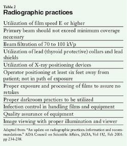
Further recommendations made in 2001 by the American Academy of Oral and Maxillofacial Radiology, ad hoc Committee on Parameters of Care, include: utilization of the paralleling technique for film exposure, proper collimation utilizing beam restricting alignment devices, and proper operator and patient barriers. In addition to utilizing the lowest amount of exposures for diagnostic purposes and the proper film speed, equipment should be checked on a daily, monthly, and yearly basis. Federal regulatory agencies require that X-ray equipment be tested every five years, depending on the state of practice. Lead shields with a thyroid collar should be utilized and stored properly unfolded when not in use. Newer lead-free shields are also available from the DUX Dental Corporation (Van R, Clive Craig, Cadco). Utilizing aerospace technology, it has thin alloy sheeting with equivalent protection against scatter radiation, but it is 50 percent lighter. This is another option for patient protection. Complete infection control protocols should be maintained to prevent cross contamination (see Table 2).
Quality control issues stem from proper exposure and the techniques utilized in processing the films. To ensure consistently high quality images, recommendations have been made to monitor and assess practitioner reliability and equipment maintenance. Studies have shown that poor diagnostic quality of radiographs from improper processing techniques can lead to higher exposure of patients. To eliminate these trends, the guidelines have been recommended. It is suggested that reference films be posted as a guide for the practitioner. A retake log should be generated to indicate the number of films exposed, along with the operator's name. If retakes are needed, these should also be noted, along with the reasons for the retakes and the corrections that were instituted. View boxes with proper illumination are to be utilized for reading the radiographs and should be checked for proper function. They should be cleaned and disinfected regularly. Proper conditions should be maintained for viewing all radiographs to ensure diagnostic quality. Darkrooms should be checked on a daily and monthly basis. Timers and chemistry should be checked monthly to assess accuracy (see Table 3).
In questioning Dr. William Beckner, director of the NCRP, about the maximum number of X-ray studies an individual can have, he stated, "There isn't a maximum number. All diagnostic studies should have a good clinical justification." Furthermore, when questioned on the minimal number of X-rays a patient should be exposed to for treatment, he stated,"The number of X-rays to be taken should be justified by weighing the expected benefit against the estimated risk, and the expected benefit should outweigh the risk. Taking X-rays on asymptomatic patients on a routine basis doesn't look at either the benefit or the risk." Perhaps routine X-rays should not be taken.
Finally, the last recommendation by the American Dental Association was continuing education. New staff should be educated on the office's policy and procedure for radiological examinations and documentation. Current staff should be updated on new processing techniques, new film speeds, and any other changes in technique or office standards. As there are always changes in new technology, practitioners should keep informed of new information.
By utilizing our expertise, we follow the ALARA principle. We must continue to use our professional judgment in exposing our patients. The benefits must outweigh any risks of radiation exposure to the patient. Radiographs are the necessary key to excellent patient care through good clinical practice and proper diagnosis. Let's use them prudently.
The author wishes to thank Dr. Harold Rabin for his invaluable assistance.
Sheri B. Doniger, DDS, practices in Lincolnwood, Ill. She graduated from the University of Illinois College of Dentistry in 1983 and obtained her bachelor's degree in dental hygiene from Loyola University of Chicago in 1976. She can be reached at (847) 677-1101 or [email protected].
References• Goaz, White, SC. "Oral Radiology", Mosby Year Book. Third Edition, 1994, 73-78.
• "The Selection of Patients for X-Ray Examinations: Dental Radiographic Examinations" HHS Publication FDA 88-8273.
• Parameters of radiologic care: An official report of the American Academy of Oral and Maxillofacial Radiology" White, SC, Heslop, E., Hollender, L., Mosier, K., Ruprecht, A., Shrout, M. Oral Surgery Oral Medicine Oral Pathology Oral Radiology Endodontics, 2001; 91: 498-511.,
• "Recommendations for quality assurance in dental radiography" Preece, J, Ed. Oral Surgery, Oral Medicine and Oral Pathology 1983, April, Vol 55: 421-426.
• "Radiation dose-related techniques in North American dental schools" Geist, J, Kats, J. Oral Surgery Oral Medicine Oral Pathology Oral Radiology Endodontics. 2002; 93: 496-505.
• "A comparison of Kodak Ultraspeed and Ektaspeed Plus dental X-ray films for the detection of dental caries" Wong, A, Monsour, PA, Moule, AJ, Bashford, KE. Australian Dental Journal. 2002;47:1: 27-29.
• "Dental Digital Radiologic Imaging" Mauriello, S, Platin, E. Journal of Dental Hygiene, Fall 2001, Vol 75, Issue IV; 323-334.
• "Dental Digital Radiography" Moore, W. Texas Dental Journal, May 2002: 404-412.
• "An introduction to digital radiography in dentistry" Brennan, J. Journal of Orthodontics, 2002, Vol. 29: 66-69.


