Nancy W. Burkhart,BSDH, EdD
What is Heck’s disease?
Human papillomavirus (HPV) infection is often associated with oropharyngeal cancer known to be correlated with the HPV 16 virus. Other HPV types are also associated with head and neck cancers. However, there are many types of low-risk HPV viruses that cause various disease states and manifestations within the mouth and elsewhere. The low-risk varieties are not associated with a malignant outcome.
Benign HPV subtypes cause the common wart, multifocal epithelial hyperplasia (MEH), condyloma acuminatum, and squamous papilloma. The high-risk types of HPV are responsible for the majority of HPV-induced cancers, while other subtypes in this category have been identified as candidates for oncogenic potential.
MEH, also called Heck’s disease, was first noted in the literature in March 1881, in groups of children of Indian descent. In 1961 Heck came across the condition again; thus, today’s name of Heck’s disease.1 MEH has reportedly been found in certain ethnic groups in areas of Mexico and in Native American groups, with a lower incidence reported in other groups such as Asians.2 A 2017 report from Ledesma-Montes and Mendez-Mendoza reports a high rate of MEH in a population of children in Mexico. The lesions in this group were commonly seen on the buccal mucosa and tongue, and a higher rate in the female population was noted.3
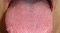
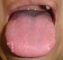
Figure 1: Multifocal epithelial hyperplasia of the right lateral border of the tongue
It is thought that there is an element of susceptibility within certain ethnicities, or perhaps crowded conditions in these groups make spread of the disease more common. MEH tends to occur in young age groups, usually within the first two decades of life, and especially in those located in close quarters, such as daycare facilities or crowded living areas. Other etiologies suggested are poor hygiene, immune suppression, genetic predisposition, and malnutrition.
MEH may also be seen in more mature patients, includingthe very old. One report discusses the case of a 92-year-old patient diagnosed with MEH who reportedly had no major health issues other than possibly his age as a contributor to low immune function, with low natural killer cell function as a factor. The authors call attention to the characteristic features of MEH and suggest that it be considered in a diagnosis even when the patient is much older than the age range normally diagnosed.4
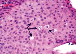
Figure 2: Histology of multifocal epithelial hyperplasia with arrows pointing to the mitotic figures (mitosoid cells) commonly found in this type of lesion. Koilocytosis associated with HPV is also found in MEH.
More facts about Heck’s disease
Names given to the disease in the literature are Heck’s disease, multifocal papilloma virus epithelial hyperplasia, focal epithelial hyperplasia, and verrucae of the oral cavity.5 The accepted name today is MEH.
Clinicians may be confused about these MEH lesions because they are multiple in number, coalesce, and exhibit a noninflammatory appearance, yet may have a slightly granular to papillary surface. The papules are rounded, smooth, or pebbly on the surface, and are firm without any exudate and have no inflammation of the papule or surrounding area. One study mentions that MEH may be found in two forms, papulonodular (pink and smooth) and papillomatous (white and pink with a pebbly surface).6
Figures 1–3 depict multifocal epithelial hyperplasia. The actual growth is usually 1 mm to 10 mm, but because the papules often coalesce, they may appear larger. The term multifocal is used because they most often occur at multiple sites as variable numbers of papules (as seen in figure 1) along the lateral border of the tongue. The papules are circumscribed, soft, and flat.
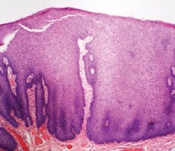
Figure 3: Acanthosis with broad rete ridges that are elongated, denoting a papillary surface of the epithelium.
The MEH data suggests a life span of anywhere from a few months to many years, depending upon the patient. The papules are usually not treated unless they are in areas of friction or trauma. Lasers, cryotherapy, or electrocoagulation are mentioned as possible treatment modalities.
A study by Agnew et al. mentions possibilities to consider or rule out when papules are observed in children, such as condyloma acuminatum, multiple endocrine neoplasia syndrome type III, neurofibromatosis, tuberous sclerosis, epidermal nevus syndrome, and Cowden syndrome.3
As always, remember to listen to your patients and ask good questions!References
1. Deliverska EG, Dencheva M. Rare case of multifocal epithelial hyperplasia of oral mucosa. J of IMAB. 2013;19(4):392-395.
2. Kubiak M, Pawe S. Focal epithelial hyperplasia (Heck’s disease) – case report of a rare disease in an adult Caucasian man. Dent Med Probl. 2015;52(4):516-520. doi:10.17219/dmp/58591.
3. Ledesma-Montes C, Mendez-Mendoza A. Unusually high incidence of multifocal epithelial hyperplasia in children of the Nahuatl population of Mexico. Indian J Dermatol Venereol Leprol. 2017;83(6):663-666.
4. Shamloo N, Mortazavi H, Taghavi N, Baharvand M. Multifocal epithelial hyperplasia: a forgotten condition in the elderly. Gen Dent. 2016;64(5):72-74.
5. Agnew C, Alexander S, Prabhu N. Multifocal epithelial hyperplasia. J Dent Child (Chic). 2017;84(1):47-49.
6. Neville BW, Damm DD, Allen CM, Chi AC. Oral and Maxillofacial Pathology. 4th ed. St. Louis, MO: Elsevier; 2016.
NANCY W. BURKHART, EdD, BSDH, AFAAOM, is an adjunct associate professor in the Department of Periodontics-Stomatology, College of Dentistry, Texas A&M University, Dallas, Texas. Dr. Burkhart is founder and cohost of the International Oral Lichen Planus Support Group and coauthor of General and Oral Pathology for the Dental Hygienist. Dr. Burkhart was awarded an affiliate fellow status in the American Academy of Oral Medicine in 2016 and Pemphigoid Foundation and is a 2017 RDH/Sunstar Award of Distinction recipient. Contact her at [email protected].


