Chairside saliva testing changes the definition of ‘restore’
by Shirley Gutkowski, RDH, BSDH, FACE
There are two kinds of language, dead and alive. With the former, new words, such as in Latin, are never added. English is certainly alive, with over 1,000 new words added every year. Words like Webinar, and staycation are combinations of older words. Some words are totally new, like muggle. And others have totally new meanings, like friending, unfriending, and tweet. Medico/dento language is alive too. Some changes make communication easier. Sometimes a word change makes communication a little more murky; it’s important to change terminology for a good reason.
The vocabulary problem that dentistry and medicine share is the use of diminutive terms and expressions. Malcolm Gladwell illustrates the problem with diminutive language in his book Outliers. The entire book is an illustration of how things are not exactly as they seem. In one chapter, Gladwell itemizes the sequence of events leading up to airline accidents. It turns out the reason is mainly due to diminutive language.
Gladwell explains, for example, that an average of seven miscommunications occur in the cockpit before the plane crashes — seven! This kind of crash is often reported as pilot error. What we don’t find out is what pilot error really means. Did the pilot flick a switch in error that made the plane fall? Did the pilot fall asleep? Did the pilot forget he was driving a plane and try to parallel park? Hardly. It’s due to a cluster of vague language between those at the helm. “Boy, it’s icy out there today,” as opposed to “There’s ice on the wings, Captain.” Really!
In our dental world, the word little is the most damaging. We use it all the time to keep our patients from totally freaking out and needing a straitjacket (that’s called hyperbole). While describing a periodontal infection, oral cancer, or enamel breakdown, using imprecise language intended to soften the news hurts our patients and the practice’s bottom line.
Can you see how this happens in dentistry? “I see a little bleeding” or “I found a little stick” are examples of diminutive speech. “Your tissue is bleeding” or “This tooth is decaying” are examples of more direct language.
The progress of change
This change in cleaning up the language for our profession may start with the term restoration. Currently, restoration is used as a noun referring to the end product of replacing tooth anatomy with a biologically acceptable material to approximate the tooth’s original dimensions, or to modify the original proportions of the tooth in some way. The verb form of the word is “restore” -- to bring something back to its original condition. People restore furniture.
A dentist is not often referred to as a restoration specialist. He or she may be thought of as a restorative dentist. However, the term refers again to the end point of a tooth exhibiting an approximation of its original size and shape.
The dental hygienist is more accurately the restoration specialist. Restoration can refer to the action of restoring the mouth to health — establishing homeostasis in the oral cavity, potentially eliminating the need for drillin’ ’n’ fillin’. Over the last decade, dentistry has narrowed the reasons enamel breaks down. Acid is the real culprit. The source of acid springs from either diet or bacterial metabolism. Studies in the late 1960s using gnotobiotic (germ-free) rats showed that the rats’ teeth would not break down without the presence of Streptococcus mutans.
Blaming sugar for all the ills associated with dental decay diverts treatment. The studies of the 1960s were very clear on that fact. What has become clearer is that the presence of sugar causes Strep mutans to pump out the sticky substance that protects the biofilm, and that the sugar metabolizes in the bacteria to produce acid.
Figure 1: Even with loupes, fissures like this can subjectively be treated using surgical means, medical means, or just waiting for change. How can a person tell if there’s been change?
The acid created affects the ecology of the biofilm, favoring acidophilic bacteria, which crowds out the basophilic (healthier) bacteria. The acid also breaks enamel bonds, which presents as a cavity in need of a restoration. If dental professionals go back a link or two in the chain of events that precedes enamel breakdown, we can confidently restore the health of the biofilm. Doing so separates the links leading to enamel decay.
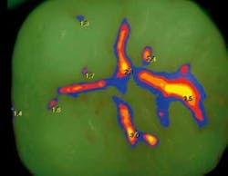
Figure 2: The red is the wavelength of Strep under Spectra’s wavelength. In this view, healthy tissue is green. Notice that some of the fissures are just stained. This image can guide a patient and dental hygienist in better home care.
Detecting the presence of oral biofilm is easy. Verifying the inhabitants of the biofilm is something else again. Most methods require expertise, equipment, and time. As industry scientists take information and refine it, dentistry’s goals for objective data to detect problems earlier are easier to achieve.
The development of chairside saliva testing has made evaluation of this very important bodily fluid much easier. Dental hygienists can evaluate the quality of saliva. We cannot yet tell if the Strep in saliva is mutans, sobrinus, oralis, or any other strain. The use of probiotics makes this distinction between bacterial strains even more important.
One of the lesser known and very accurate ways of detecting bacterial activity is by using fluorescence. Fluorescence is a simple process of using a predetermined wavelength of light to shine on a sample of biological material — in this case, an oral biofilm — and measuring the reflected wavelength off of the material. Everything in the world has signature fluorescence. If it sounds difficult or New Age, consider that fluorescence has been around in dentistry for nearly a decade, as well as for multiple decades in science and industry.
Practical application of fluorescence in dentistry has increased. Air Techniques, for example, has developed an uncomplicated way to gather data with their Spectra Caries Detection Aid. The wandlike handle of the Spectra allows the clinician to move thoroughout the mouth and read the fluorescence of the oral biofilm on each tooth, much like taking intraoral images.
Visualizing the type of biofilm is very liberating. Figure 1 is an actual view of the occlusal surface of a premolar. The pits located at 8 o’clock position appear to be stained. Many experienced dentists confidently leave that type of fissure undisturbed or treat it by amputating the darkened enamel, or “watch it.” Even with loupes, there is no ability to make a definitive treatment plan without help.
Figure 2 shows the same tooth under the wavelength used by the Spectra. Most of the tooth reflects the color back to the receptor. The pits and fissures, however, reflect a different quality of green and even some red (red material in the dental world is viewed as cariogenic biofilm).
Figure 3: This image shows how the software relays information to the clinician. In the continuum of colors, blue is to the far left as healthy. Red to orange to yellow shows a progression of enamel breakdown starting with surface damage down to dental and pulpal involvement. The numbers give the clinician another level of objective information where they do not have to judge a color’s value.
Figure 3 reveals another feature of the Spectra software. This setting allows the clinician to detect the difference in enamel density as affected by the cariogenic bacteria. For instance, the blue enamel is relatively sound, the red reveals enamel breakdown, the orange reveals compromised dentin, and the yellow lets the clinician know that the damage has entered far into the dentin. The Spectra software also gives the color a numeric value, providing the clinician ample information to make a diagnosis, plan a treatment, and make a prognosis as to the potential outcome of the prosthesis should one be necessary.
The dental hygienist or the dental assistant can take the images as data collection before the dentist even sees the patient. This kind of information is vital in determining treatment plans.
With the screen above the patient, in this photo, the clinician can easily discuss Spectra findings. The mass of red is a biofilm of cariogenic bacteria on the side of the tooth. The blue shows currently healthy enamel.
This entire suite of information is then part of the patient’s electronic record, just like digital X-rays. There’s no second step to write everything out or type into the office software. Sequential images of the same tooth can then be lined up side by side to show disease progression or healing. The Spectra hardware can integrate with any computer and is compliant with dental imaging and practice management software programs. There’s no need to have multiple laptop computers in each room. More importantly, the patient can be more involved than ever before.
Dr. Peter Wilt uses advanced technologies in his Hartford, Wis., dental practice. Kim Grensavitch, RDH, works in the practice as well and finds using technology including caries/biofilm detection with Spectra makes treatment of patients’ conditions easier. Dr. Wilt does a different kind of dentistry using Spectra. The discovery of early enamel breakdown allows him and his team to treat lesions earlier, amputate less of the tooth, and have better outcomes. Kim is able to track the changes of her white spot treatments using objective technologies.
“The state of the art is always changing,” says Wilt. “A dentist can certainly still do good dentistry without these advanced technologies; with them, dentistry is better, easier, and I can be more confident in the outcomes.”
Dr. Wilt uses the technology as he removes diseased enamel to help him determine if more tissue needs to be removed. This simple process of stopping to take an image gives him confidence in his clinical decision-making.
Other fluorescence tools give similar information. Fluorescence along with measuring the health of the saliva can guide a clinician toward restoring the entire mouth to health.
Dentistry still needs a word to replace the noun restoration: prosthesis. As defined by Webster.com, a prosthesis (prosthetic) replaces or augments a missing or impaired body part. That’s what a filling or crown does. Calling a prosthesis a restoration makes it OK to need one even in the minds of dental professionals when, in fact, it should be a sad turn of events. The industry’s tolerance of such surgery will decrease by replacing diminutive language with more accurate verbiage.
Look for other ways to refine or redefine the language of dentistry as industry supports us with instruments and medicines to restore oral health.
The notion of going backward is very difficult for forward-thinking people, but moving purposefully means looking for opportunities for taking corrective measures.
The history of diminutive or imprecise language comes from dentistry’s early days. Dentistry was brutal. In order to have patients understand or even return for their next visit, it was easier, even healthier, for them to think that the problem presented was small. In those days, patients had to rely on the word of the dentist. That’s not so today. Patients have many options for co-diagnostics that rely on third-party science including a dental hygienist and the Internet.
The question then becomes: What words need replacing? Or more accurately: What words can we use to make sure our patients understand what’s going on, don’t get freaked out, and actually schedule dental care?
The answers may come from knowing and understanding what instruments and medicines are available to help us make a real diagnosis (as compared to a determination) and engage our patients in the discovery phase of diagnosis.
Shirley Gutkowski, RDH, BSDH, FACE, is Technology Director of CareerFusion and a practicing dental hygienist. She is author of The Purple Guide: Confronting Caries, available at www.rdhpurpleguide.com. The author can be reached at [email protected].


